


We often picture the back as a stack of vertebrae separated by discs. Biotensegrity offers another view: bones, muscles, ligaments, and fascia work together as a coupled system that can transmit and redistribute mechanical loads. Recent studies now make it possible to measure some of these remote mechanical interactions in living humans.
A note before reading. Biotensegrity is a biomechanical model. It does not mean that fascia alone explains back pain, nor that tension located at a distance automatically identifies where pain comes from. The data presented here are meant to help you better understand how the system works mechanically.
The Dinosaur Skeleton That Changed How We See the Body
In the mid-1970s, American orthopedic surgeon Stephen Levin was looking for a model that could explain the stability and movement of the skeleton as something more than a simple assembly of levers. Levin recalls that he regularly visited the Smithsonian National Museum of Natural History to study dinosaur skeletons, yet could not make their architecture fit the classical mechanical model he knew. [1]
On the National Mall in Washington, another structure caught his attention: Kenneth Snelson’s Needle Tower, installed at the Hirshhorn Museum, part of the Smithsonian. In this sculpture, the rigid elements do not rest on one another: they are held in place by a continuous network of cables under tension. [1] [2]
Snelson had developed this principle of “floating compression” after working with architect R. Buckminster Fuller. It was Fuller who coined the term tensegrity, a contraction of tensional integrity, to describe this combination of continuous tension and discontinuous compression. [2]
The bars don’t stack. And yet, the whole structure stands.
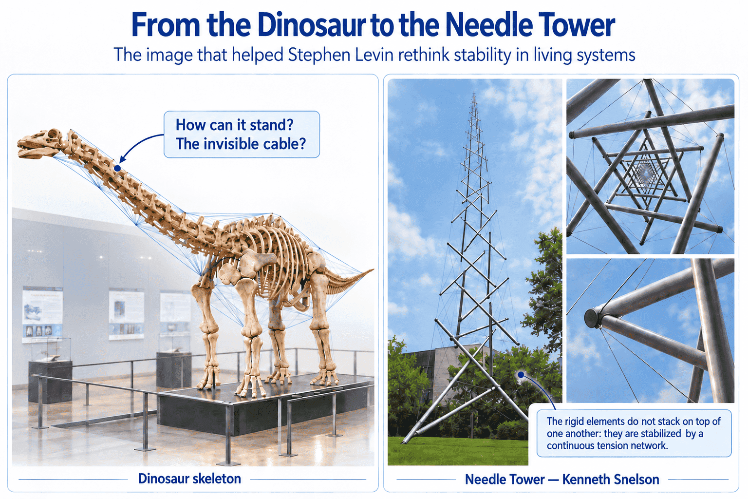
Levin saw in this architecture a different way of thinking about living systems. He would go on to develop the concept of biotensegrity: the application of tensegrity principles to biological organization, particularly to the spine. [3]
A Spine Is Not a Tower of Blocks
We tend to imagine our skeleton as the frame of a building. The vertebrae would be stacked on top of one another, the discs would act as shock absorbers, and the muscles would pull on the whole structure to produce movement.
This picture is useful.
However, it is incomplete.
In the biotensegrity model proposed by Levin, biological structures are viewed as systems in which elements under compression are integrated into a network of distributed tension. Applied to the spine, this model leads us to consider bones, muscles, ligaments, fascia, and joints together rather than isolating each part. [3]
A vertebra never works alone. A muscle never pulls into empty space.
The Spine: A Spider’s Web, Not a Tower of Blocks
This is therefore where fascia becomes particularly interesting.
Fascia is connective tissue organized in networks and layers, associated with muscles and many other structures. The current literature describes its role in transmitting mechanical loads and in interactions between anatomically connected regions. [4]
However, this does not mean that “everything comes from fascia.”
It means something more interesting:
A mechanical load is not necessarily confined to the place where it appears. Some interactions can be transmitted through connected musculofascial structures. [4]
For a long time, this idea relied mainly on anatomy, biomechanical models, and indirect experiments.
Now, it is becoming possible to observe some of these interactions in a living human being.
Why It Changes Everything: Proof Through Measurement
In 2024, Eleftherios Kellis, Afxentios Kekelekis, and Eleni Drakonaki published a particularly interesting study in the Journal of Anatomy.
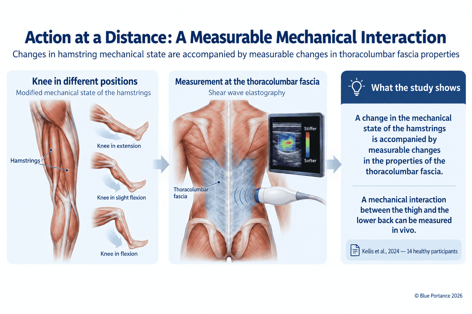
The researchers examined 14 young, healthy men. They used shear-wave ultrasound — an elastography technique that estimates the mechanical properties of a tissue — and simultaneously measured the thoracolumbar fascia and the fascia of the semitendinosus and semimembranosus muscles. [5]
The protocol is simple: the knee is placed at 0°, 45°, and 90°, first passively, then during submaximal isometric contractions of the knee flexors. [5]
So What Happens?
When the knee moves passively from 90° of flexion to extension, the shear modulus of the thoracolumbar fascia increases significantly. A significant increase is also observed during active contraction of the knee flexors. [5]
In other words, you act at the level of the thigh, and a mechanical change becomes measurable in the tissues of the lower back.
The authors conclude that hamstring exercise may remotely influence the stiffness of the fascia surrounding the lumbar region. [5]
However, this study does not show that hamstring tension causes low back pain. It shows something more precise: a remote mechanical interaction can be measured in vivo under the conditions studied. [5]
An Old Thigh Injury… and Different Lower-Back Mechanics
In addition, the same team took the observation a step further in a second study published in 2024.
It compared 11 soccer players with a history of hamstring injury with 13 players without a comparable history. The properties of the thoracolumbar fascia, the erector spinae, and the multifidus were measured using the same elastography technique. [6]
The modulus of the thoracolumbar fascia was significantly higher in the group with a history of injury. The authors also observed higher values for some lumbar muscles. [6]
Still, we should be careful not to jump to conclusions: this cross-sectional study does not show that the thigh injury caused this difference in the lower back. It highlights an association between a history of distal injury and different mechanical properties in the thoracolumbar region. [6]
The nuance is essential: a remote mechanical relationship can be measured without turning that relationship into a diagnosis of causality.
So, Can Tension in the Leg Cause Back Pain?
This is where we need to resist an appealing but overly simple conclusion.
The studies above do not allow us to claim: “your low back pain comes from your hamstring.”
Likewise, they do not show that tension in the foot or calf automatically travels up to the lower back.
On the other hand, they show that remote mechanical interactions exist and can be objectively measured.
A systematic review published in 2025 identified 19 studies meeting its inclusion criteria on remote myofascial interventions. The included studies report effects on range of motion, pain, or function, among others, but the results are heterogeneous and the authors emphasize how difficult it is to isolate a single mechanism. [4]
The review discusses several possible explanations, including mechanical transmission through tissues and neurophysiological mechanisms. Their respective contributions have not been definitively settled. [4]
And that is precisely what makes a living body more interesting than an assembly of parts.
So Where Does Back Pain Fit In?
This is where biotensegrity becomes truly useful.
Not because it provides a new structure to blame.
But above all because it changes the question.
The classic question is:
“Which part is damaged?”
The disc? A vertebra? A joint? A muscle?
These questions remain important. However, a systems-based reading lets us add others:
- how are the loads distributed?
- which movements are still available?
- can the pelvis and hips easily change configuration?
- do the tissues still have room to glide and deform?
- are some regions compensating for others over the long term?
We then shift from a parts-based logic to a systems-based logic.
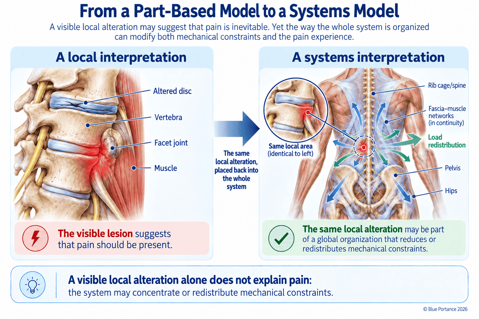
This approach also helps explain why imaging does not always tell the whole story of back pain. This topic is covered separately in the article: Back Pain with a Normal MRI: Why Do You Still Hurt?
The key idea here is different: the absence of a local lesion that explains the pain does not mean there is no mechanical organization, compensation, or interaction between structures.
The Real Issue: Being Able to Redistribute
Imagine two systems subjected to the same load.
The first has many options: it can slightly shift the pelvis, change the weight-bearing on one hip, adjust muscle tension, vary the areas under load, and reorganize its movement.
By contrast, the second has only a few solutions left.
As a result, it repeats the same organization over and over again.
The fundamental difference between the two may not be just their posture. It is their ability to adapt.
From Biotensegrity to Adaptability
This is precisely where this reading connects with the SBNFA™ — Systemic Biological Neuro-fascial Adaptive Framework.
In the SBNFA™, the analysis does not focus solely on a position or an isolated structure. It looks at how the system maintains — or loses — its capacity for adjustment.
In particular, the reading focuses on the ability to:
- vary points of support;
- preserve degrees of freedom;
- redistribute loads;
- engage body segments in different ways;
- allow tissues to glide and deform;
- alternate strategies over time.
This part belongs to the SBNFA™ framework: it is a functional reading developed by Blue Portance from a body of biomechanical, neurophysiological, and tissue data; it should not be confused with a direct conclusion of the Kellis studies.
The right question is no longer just “what is your position?” but also “how many options do you still have to change it?”
And When We Sit?
Finally, sitting makes this question especially concrete.
For several hours, the pelvis remains in contact with an external surface. When the possibilities for movement are reduced, the same areas may remain under load for a long time.
The goal, then, is not only to seek a “good posture,” but to keep enough freedom to:
- change points of support regularly;
- move the hips;
- allow the pelvis to change orientation;
- avoid holding the same mechanical configuration for too long.
In the SBNFA™ reading, stability is therefore not necessarily synonymous with immobility: a system can remain stable while retaining small adjustments and several response strategies.
This reversal is what guided the design of the dynamic Aporia® seat. For decades, seating ergonomics has sought to hold the body in a “good posture”: support it, wedge it in, stabilize it. Aporia® starts from the opposite principle. Its patented technology does not lock the pelvis in place: it restores mobility within stability, allowing micro-adjustments, varied points of support, and hip movement over time (see Figure 4).
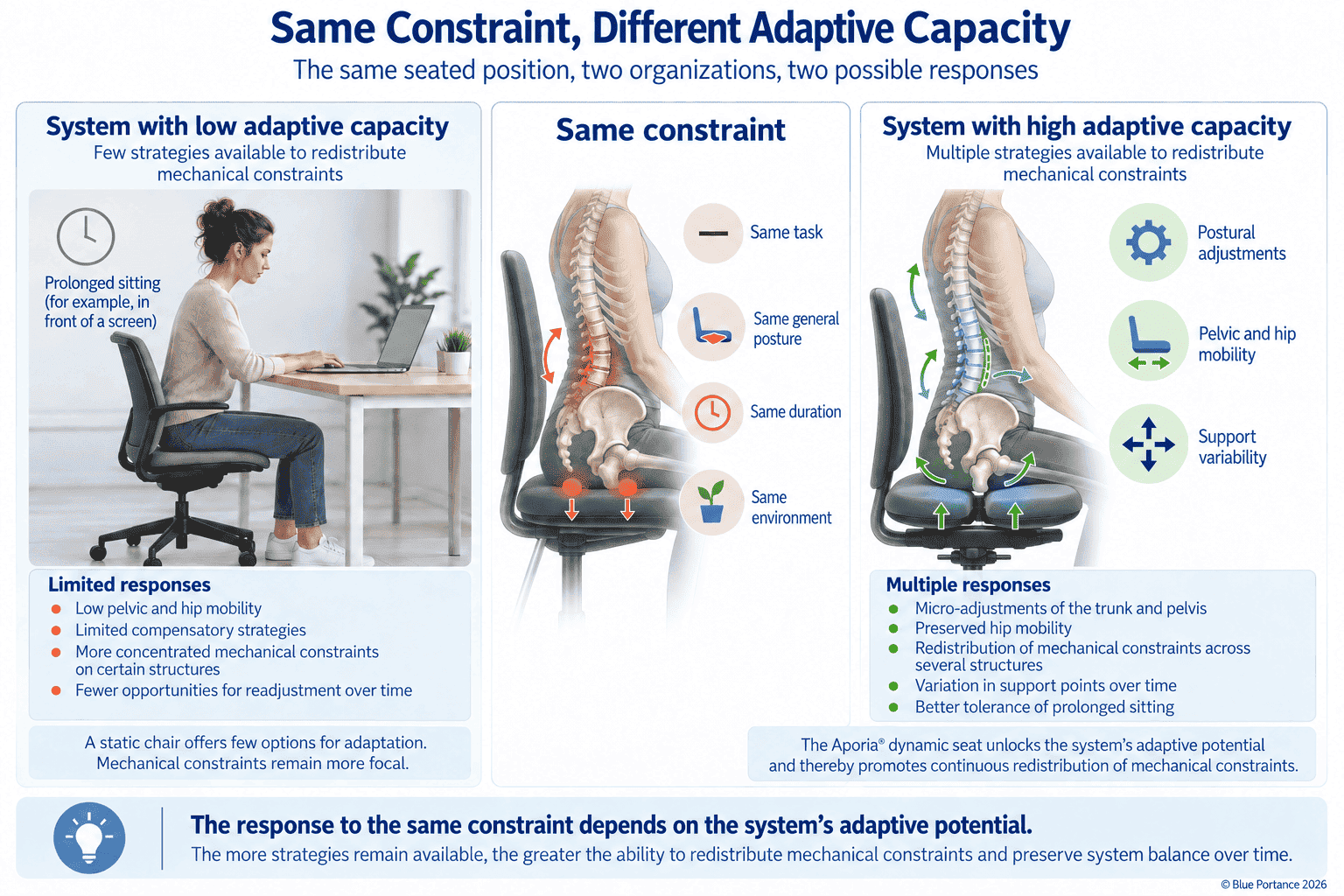
The seat no longer corrects a posture. It unlocks the system’s full adaptive potential.
Conclusion: A Back Is Not a Column — It’s a Living System
Biotensegrity does not replace anatomy, imaging, or medical diagnosis.
Instead, it brings another scale of understanding.
Recent research shows that some mechanical interactions between distant regions can be measured in humans [5] [6], while the mechanisms linking these interactions to symptoms are still partly debated. [4]
To understand back pain, this invites us to look not only at what is damaged, but also at what can still move, redistribute, and adapt.
Because a back is not just a spinal column.
It is a living system.
Further Reading
-
Back Pain with a Normal MRI: Why Do You Still Hurt?
To understand why the intensity of pain does not always match the abnormalities visible on imaging. -
Fascia and Pain: Understanding Tensegrity and Load Regulation
To explore how fascia is organized, how tissues glide, and how loads are transmitted. -
Adaptability and Adaptive Potential
To understand why the ability to change configuration is a central dimension of functional analysis.
Sources and References
- Stephen M. Levin — account of how his thinking on biotensegrity began. The Levin Biotensegrity Archive and the archives devoted to his work describe his visits to the Smithsonian in the 1970s and the triggering role of Kenneth Snelson’s Needle Tower. View source.
- Smithsonian Institution — Kenneth Snelson, the Needle Tower, and the origin of the term “tensegrity.” The Smithsonian notes that Snelson developed the principle of floating compression and that Buckminster Fuller coined the term “tensegrity.” View source.
- Levin SM. The Tensegrity-Truss as a Model for Spine Mechanics: Biotensegrity. Journal of Mechanics in Medicine and Biology. 2002;2:375–388. DOI: 10.1142/S0219519402000472.
- Jafari B, Minoojejad H, Sheikhhoseini R, Rajabi R. Effect of remote myofascial intervention on musculoskeletal health and functional performance: a systematic review. Advances in Rehabilitation. 2025;39(2). View article.
- Kellis E, Kekelekis A, Drakonaki EE. Is thoracolumbar fascia shear-wave modulus affected by active and passive knee flexion? Journal of Anatomy. 2024;244(3):438–447. DOI: 10.1111/joa.13977. PubMed.
- Kellis E, Kekelekis A, Drakonaki EE. Thoracolumbar Fascia and Lumbar Muscle Stiffness in Athletes with A History of Hamstring Injury. Journal of Sports Science & Medicine. 2024;23(2):436–444. DOI: 10.52082/jssm.2024.436. PubMed.
