

Why knee pain can originate from the pelvis — and how your brain sometimes misinterprets these signals?
Summary — Fascia is not just connective tissue. It is a dynamic, body-wide network that distributes mechanical forces, enables tissue gliding, and continuously communicates with the nervous system. When this system loses adaptability — especially during prolonged sitting — forces concentrate, gliding decreases, and pain can emerge even without visible tissue damage.
Fascia: the missing link between movement and pain
Fascia and pain: Do you experience pain when sitting… yet your medical exams are normal?
Did you know that your body contains an invisible network — a system embedded within connective tissue, representing approximately 20 to 30% of total body mass, connecting every structure in your body?
This network is known as fascia, long considered merely anatomical “wrapping” with no major functional role.
Today, research in biomechanics and neuroscience reveals that fascia forms a highly active, adaptive and reactive system, essential for mobility, stability, mechanical force distribution, and even pain perception.
Understanding the relationship between fascia and pain is key — especially in prolonged sitting.
To fully understand its importance, let’s explore its structure, functions, and mechanisms step by step.
1. Fascia: the invisible network connecting your entire body
Fascia plays a central role in how mechanical stress is distributed in the body and in the emergence of pain, especially in prolonged sitting. To understand how fascia regulates mechanical forces in the body and interacts with the neurosensory system, we must first examine its structure — how it is organized and what it is made of.
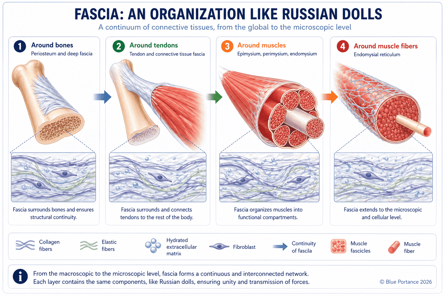
1.1. A continuous, nested structure throughout the body
Fascia are connective tissues forming a continuous network throughout the body. Unlike a segmented anatomical view where each structure is independent, fascia creates a true anatomical continuum, linking all structures together:
- Muscles (and their surrounding layers)
- Bones and joints
- Organs (heart, lungs, digestive system…)
- Nerves and blood vessels
Fascia are not limited to an outer layer. They exist at every level: superficial (just beneath the skin), between muscles, around organs, and deep within internal structures. This organization means no part of the body is truly isolated.
Fascia is organized into successive layers, interconnected, forming an architecture that is both continuous (without interruption) and hierarchical (from superficial to deep layers).
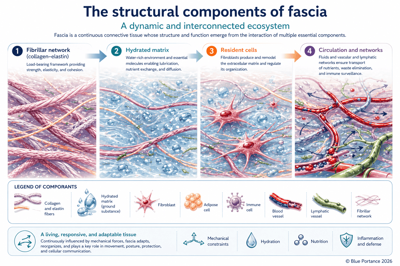
1.2. The fundamental components of fascia
To understand how fascia regulates mechanical forces in the body — and why dysfunction can lead to stiffness or pain — we must examine its internal organization. This structure is based on three key components, constantly interacting to provide flexibility, resistance, and adaptability:
A. The extracellular matrix — the lubricating “gel” enabling sliding
The extracellular matrix is the environment in which fibers and cells are embedded. Composed mainly of water (60–70%), hyaluronic acid, and proteoglycans, it forms a viscoelastic substance — both fluid and resistant.
Its role is essential:
- Lubrication of fascial layers to allow optimal tissue gliding
- Shock absorption thanks to its viscosity
- Cell nourishment through nutrient and waste exchange
Imagine two glass plates separated by a thin layer of oil. When lubrication is present, they slide smoothly. If the oil dries out, friction appears. This is exactly what happens in fascia: proper hydration enables fluid movement, while dehydration leads to stiffness and pain.
B. Fibers — the adaptive structural framework
Fascia fibers, primarily composed of collagen (strength) and elastin (elasticity), form the structural framework of the tissue.
They organize dynamically:
- According to mechanical stress: fibers align and densify where forces are greatest
- With mechanical memory: prolonged postures (like sitting) can reshape their organization
Think of a fishing net: its mesh tightens under stress, and returns to shape when released. This explains fascia’s ability to both resist and adapt.
C. Cells — the system’s sensors and regulators
Fascia is not inert: it contains active cells, including:
- Fibroblasts: produce and remodel collagen and matrix
- Mechanoreceptors: detect tension, pressure, and stretch, sending signals to the nervous system
Fibroblasts act like construction workers, constantly rebuilding the tissue. Mechanoreceptors act like sensors, alerting the brain to abnormal stress.
Fascia is not a passive wrapping tissue. It is a living, adaptive system where:
- The matrix enables gliding and shock absorption
- Fibers provide structure and adaptability
- Cells regulate and communicate with the nervous system
This constant interaction explains why fascia plays a central role in mobility, stability, and pain perception.
2. Fascia: the dynamic architecture of the human body
We now know that fascia forms a continuous network connecting every part of the body. But this anatomical organization is only the foundation: the real power appears when we understand what this network actually does.
Imagine a tissue capable of:
- Instantly redistributing mechanical forces to protect joints, like an invisible safety net absorbing and spreading every load.
- Acting as a distributed sensory system, constantly communicating with the brain by sending millions of signals about posture, movement, and micro-tensions in the body.
- Continuously adapting, adjusting tension dynamically to maintain balance whether sitting, standing, or moving.
These capabilities rely on two fundamental properties that transform fascia into far more than a simple support tissue:
A. Tensegrity
A principle of dynamic balance where tension and compression interact to maintain stability without rigidity.
B. The neurosensory system
A network of sensors continuously sending information to the brain, influencing proprioception and pain perception.
Together, these mechanisms make fascia the body’s invisible regulation system — a system so powerful that when it becomes dysfunctional, it can be at the origin of unexplained chronic pain.
Let’s begin by exploring the first pillar: tensegrity, the principle that allows your body to remain stable and mobile, without relying on rigidity.
2.1. Tensegrity: stability without rigidity
Tensegrity (a contraction of tension and integrity) is an architectural principle in which elements under tension (fascia) balance elements under compression (bones). Described by Buckminster Fuller and applied to biology by Ingber (1998), it explains how the body maintains stability without excessive rigidity.
If you feel pain in the coccyx, pelvis or lower back when sitting, the issue may not come only from that area, but from how your body distributes — or fails to distribute — mechanical forces.
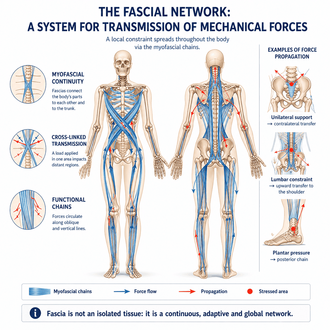
Within fascia, tensegrity enables:
- Uniform distribution of forces: tension spreads across the fascial network, preventing local overload. Example: when carrying a heavy bag, shoulder fascia redistributes load toward the pelvis and legs.
- Passive stability: unlike muscles, fascia maintains balance efficiently without fatigue. This is why you can stand without conscious effort.
- Immediate adaptability: tension adjusts continuously in response to movement. Example: when bending forward, posterior fascia tightens while anterior fascia releases.
Imagine a tent where poles (bones) resist compression while cables (fascia) maintain tension. If one cable changes, the entire structure adapts instantly. This is how the body distributes forces without overloading a single area.
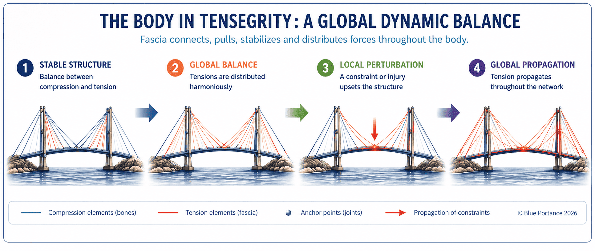
This organization operates at multiple biological scales, from cells to the entire body (Ingber, 1998). It implies that a local tension (for example in the pelvis) can produce distant effects (lumbar or cervical pain), altering global balance. This principle is central to modern therapeutic approaches such as osteopathy and myofascial chains (Myers, 2014).
Localized pain can often originate from a distant imbalance in tensegrity.
2.2. The neurosensory network
Viewing fascia as purely mechanical is like studying an electrical network without considering the current flowing through it. Fascia also forms a distributed neurosensory system, due to its high density of mechanoreceptors. As shown by Schleip (2003), this transforms fascia into a continuous communication system with the brain, influencing both proprioception and pain perception.
Fascia does not only transmit forces — it transmits information. Perception and regulation are not centralized, but emerge from continuous interaction between tissues and the nervous system.
Research by Langevin suggests that connective tissue acts as a body-wide signaling network, capable of transmitting mechanical information that influences cellular and neural behavior (Langevin, 2006). Every tension, micro-movement, or pressure change is immediately detected and transmitted.
The brain therefore receives not a single signal, but a continuous flow of information from the entire fascial network. It interprets this data to adjust posture, muscle tone and movement coordination.
A. Mechanoreceptors: ultra-sensitive sensors
Three types of receptors play a key role:
- Ruffini endings: sensitive to slow stretch and sustained tension, essential for proprioception.
- Pacinian corpuscles: responsive to vibration and rapid pressure changes, enabling real-time movement adaptation.
- Free nerve endings: detect excessive stress and can trigger pain signals.
These receptors continuously inform the brain about:
- Tissue tension state
- Body movement
- Pressure variations
When you close your eyes and touch your nose, fascia mechanoreceptors inform your brain of your arm and head position. This enables precise movement even without visual input.
B. Proprioception and pain: two sides of the same system
The fascial neurosensory network influences:
- Proprioception: your ability to perceive body position and movement.
- Nociception: your ability to detect potentially harmful stimuli.
When this system becomes dysfunctional:
- Allodynia: non-painful stimuli become painful
- Hyperalgesia: pain is amplified beyond the actual stimulus
In chronic low back pain, fascial mechanoreceptors can become hypersensitive. The brain then interprets normal tension as pain, even without structural damage.
Fascia is not just a mechanical system. It is an active interface between biomechanics and perception.
3. Fascia as an organ of regulation and harmonization of mechanical force distribution
3.1. Automatic redistribution of forces
Understanding the link between fascia and pain allows us to move beyond a purely local view of symptoms. Through tensegrity, fascia ensures a redistribution of mechanical forces that goes far beyond a simple support function. When a force is applied to the body, it is not absorbed locally, but diffused through the fascial network, allowing the harmonization of mechanical forces at a global scale.
Every movement, every posture, every effort generates forces that travel through the body. Without fascia, these forces would concentrate on specific points — joints, nerves, organs — leading to overload and tissue damage. Thanks to its continuous network organization, fascia diffuses and distributes these forces throughout the entire body.
Concrete example:
When you walk, the impact of each step is absorbed by the plantar fascia, then transmitted to the fascia of the legs, pelvis, and even the back. This redistribution system prevents shocks from concentrating on the knees or vertebrae, helping protect your joints over the long term.
This mechanism shows that stability does not rely on rigidity, but on the system’s ability to distribute and adapt mechanical forces.
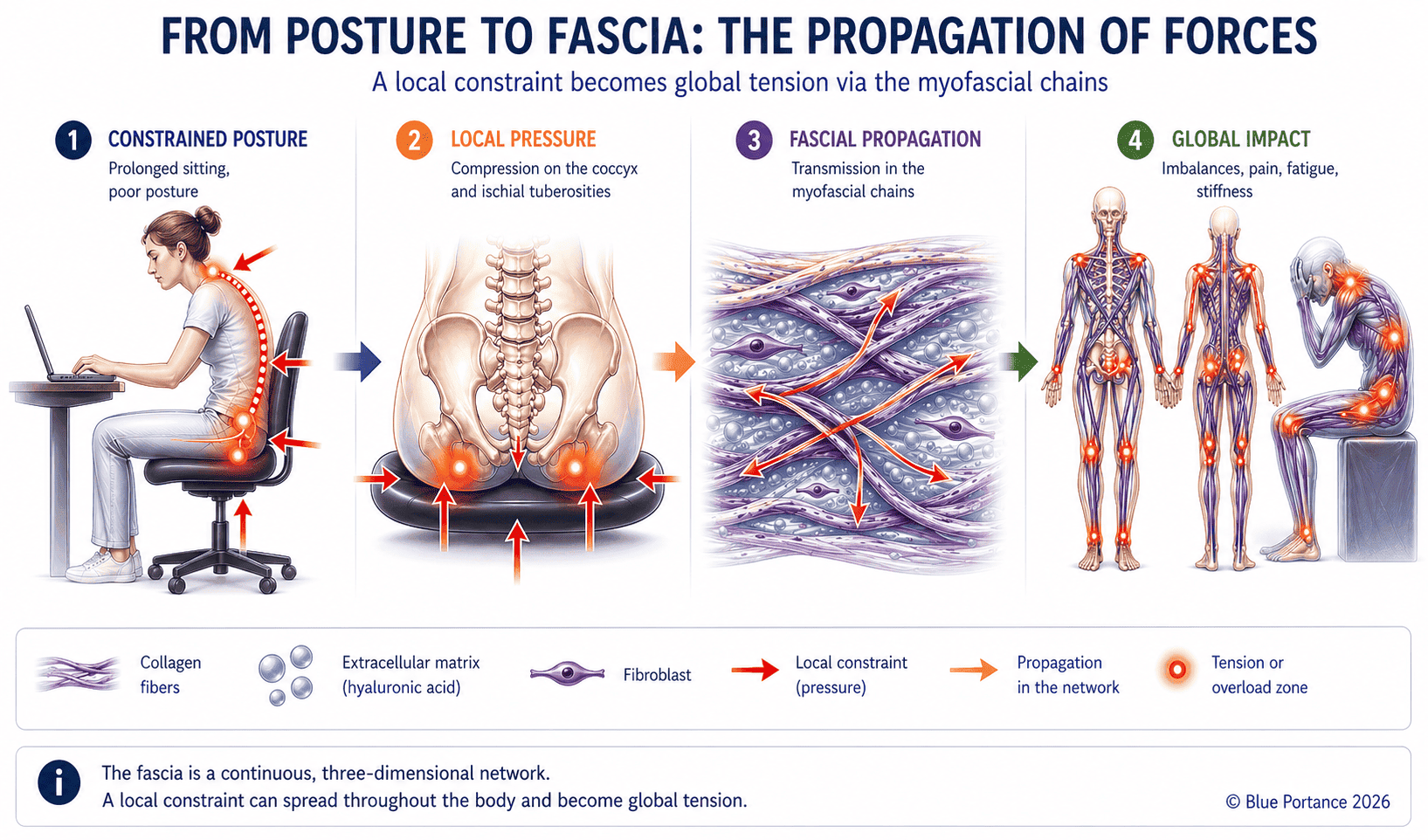
Fascia also displays viscoelastic behavior: when it is loaded, it absorbs part of the mechanical energy, but does not return all of it. A fraction of this energy is dissipated, especially as heat — a phenomenon known as hysteresis (Fung, 1993; Humphrey, 2002).
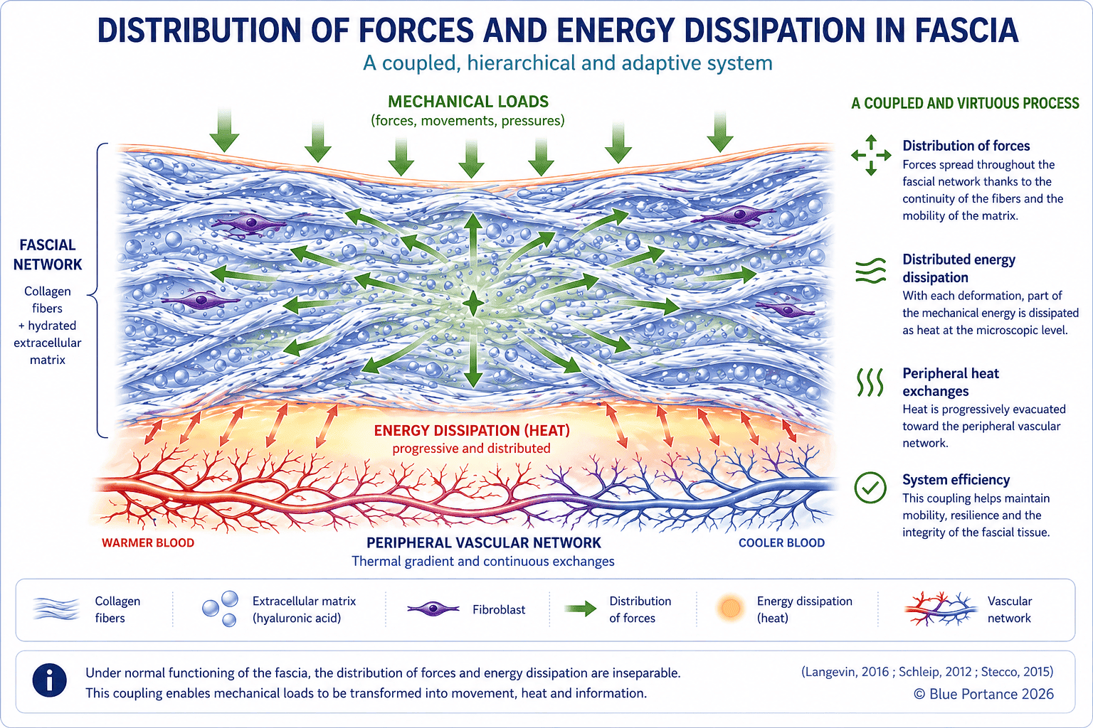
In a functional system, this dissipation remains distributed throughout the fascial network and contributes to mechanical shock absorption. However, when mechanical loads become repetitive and poorly variable — as in prolonged sitting — hysteresis increases locally. Mechanical energy no longer circulates homogeneously and tends to concentrate, reflecting a progressive loss of tissue mechanical efficiency (Langevin, 2016).
3.2. Maintaining stability without conscious effort
Unlike muscles, which require voluntary activation and become fatigued, fascia provides continuous stability through its elasticity and network organization. It enables constant adaptation to micro-variations in posture, without conscious effort.
In the sitting position, a functional pelvic fascia automatically distributes body weight between the coccyx, the ischial tuberosities, and the surrounding structures. This distribution prevents pressure from concentrating on a single area and limits the onset of pain.
When this adaptive capacity is preserved, posture remains dynamic, even in a static situation.
3.3. Protection of sensitive structures
Fascia also plays an essential role in protecting sensitive structures, especially nerves and blood vessels. It creates gliding spaces that allow these structures to move freely while cushioning mechanical stress.
In pudendal neuralgia, pain is not always caused by direct nerve compression. It may result from altered gliding within the nerve’s fascial environment, linked to a loss of force harmonization in the pelvis. This alteration changes the mechanical signals perceived by local receptors, which are then interpreted by the nervous system.
This phenomenon is supported by experimental studies showing that, in patients with chronic low back pain, the gliding capacity of fascia can be reduced by approximately 50% compared with asymptomatic subjects (Langevin et al., 2011).
This alteration of gliding is not necessarily linked to a visible lesion, but to a modification in the properties of connective tissue, including collagen densification and reduced mobility between layers (Langevin, 2016).
Pain therefore appears as the result of an interaction between local mechanics and global perception.
4. When tensegrity and force harmonization are disrupted
4.1. Causes of imbalance
Before going into detail, here is what happens when the system loses its ability to adapt.

Tensegrity can be disrupted when the mechanical properties of fascia change, particularly in situations of sedentary behavior, repetitive loading, or prolonged mechanical stress. In these conditions, the tissue gradually loses its adaptability, and forces are no longer properly redistributed across the system.
At the cellular level, this mechanical alteration is associated with changes in fibroblast behavior, the main cells of connective tissue. In response to repetitive and low-variability stress, fibroblasts can increase collagen production, contributing to fascial densification and reduced gliding between layers (Langevin, 2016).
Conversely, varied mechanical stimulation — particularly gentle stretching and micro-movements — modifies fibroblast behavior: they elongate, reorganize their cytoskeleton, and actively participate in regulating tissue tension (Langevin et al., 2005; Langevin et al., 2017).
When pelvic fascia loses its ability to adapt, mechanical forces concentrate locally. This concentration can overload sensitive structures such as the coccyx, lumbar spine, or pelvic nerves.
4.2. Consequences: pain and compensations
When tensegrity is altered, the body develops compensatory strategies. These adjustments help maintain a certain level of balance, but they can also generate new mechanical stress in other areas.
An abdominal scar can modify local fascial tension. This alteration propagates through the surrounding network, disrupting pelvic tensegrity. The body compensates, but these compensations can lead to pelvic or lower back pain, even when the original cause is no longer obvious.
These mechanisms show that system disruption does not depend only on the intensity of forces, but also on their repetition and lack of variability, which progressively alter the mechanical and biological properties of the tissue (Langevin, 2016; Stecco, 2015).
4.3. The role of the neurosensory system in chronic pain
When these disturbances persist, the neurosensory system can also change. Mechanoreceptors become more sensitive, and the nervous system may amplify incoming signals. This phenomenon is closely related to what is described as central sensitization (Latremoliere & Woolf, 2009).
In this context, pain is no longer solely linked to the initial mechanical constraint, but to how that constraint is interpreted by the nervous system. The brain may maintain or amplify pain perception even when the original mechanical cause has diminished.
4.4. Why can pain exist without visible tissue damage?
It is common to observe persistent pain even when medical imaging (MRI, CT scan, X-ray) shows no identifiable lesion. This does not mean the pain is “imaginary” — it reflects a change in how the system functions.
In a living system, pain does not depend solely on structural damage, but on the interaction between several dimensions:
- Tissue mechanics: loss of gliding or poor force distribution can create local stress not visible on imaging.
- The fascial network: altered tensegrity can disrupt global balance and create distant overload zones.
- The neurosensory system: mechanoreceptors may become hypersensitive, amplifying otherwise moderate signals.
The brain does not interpret a “lesion”, but a set of signals coming from the body. When these signals become inconsistent or amplified, it can generate pain perception even in the absence of identifiable tissue damage.
In chronic low back pain, patients may experience significant pain despite normal imaging. Studies suggest this can be linked to reduced fascial gliding and increased nervous system sensitivity, rather than visible structural injury.
Pain can be real, measurable, and disabling, even without visible tissue damage. In such cases, it reflects a dysfunction in the system regulating mechanical forces and perception, rather than a localized injury.
5. Restoring system dynamics: why micro-movements change everything
If pain does not come only from a lesion, but from an imbalance in the system — mechanical, fascial, and neurosensory — then the key question becomes: how can normal function be restored?
Contrary to common belief, it is not immobility or a “perfect static posture” that provides lasting relief. What is most often missing is mechanical variability: continuous micro-movements that allow the system to function as it was designed to.
- They restore fascial gliding by reactivating fluid exchange within the extracellular matrix.
- They redistribute mechanical forces, preventing overload on sensitive areas (coccyx, lower back, pelvic floor…).
- They reactivate the neurosensory system, sending the brain coherent and varied signals.
Conversely, a static sitting posture — even when considered “ergonomic” — promotes repetitive loading, increased local hysteresis, and a progressive loss of adaptability in the system.
It is not only the position that matters… but the ability to keep moving continuously within that position.
5.1. Why is this difficult in real life?
Because most seating systems restrict exactly what your body needs: freedom of micro-adjustment.
They provide stability… but at the cost of loss of fine mobility. As a result, the system can no longer distribute forces or regulate tension effectively.
5.2. What does this mean for you, concretely?
As long as these micro-movements are not restored, the system remains locked: mechanical stress repeats, overload zones persist… and pain continues.
This is precisely the problem that led to the development of a different approach to sitting: not to find the “perfect posture”, but to restore the body’s ability to continuously adapt.
Move from understanding to action
If you recognize yourself in these mechanisms — sitting pain, persistent discomfort, or a feeling of stiffness — then the challenge is no longer simply to understand them, but to identify what may be mechanically maintaining these patterns.
The key question is now simple: does your seat allow your body to function… or prevent it from doing so?
👉 Analyze the mechanisms involved in your sitting pain through our expert analysis system — and discover which Aporia® seating solution may best fit your situation.
6. Conclusion: the body as an orchestra
The body can be understood as an orchestra. Fascia acts both as the instruments and as the network that transmits mechanical forces and information throughout the system.
The nervous system acts as a conductor — but not an independent one. It constantly adjusts its interpretation based on the signals it receives from the body.
Modern pain neuroscience shows that pain is not a simple signal coming from tissues. It is an active process: the brain interprets, modulates, and can even anticipate signals depending on context, experience, and internal state.
This adaptive capacity relies on neuroplasticity — a fundamental mechanism that allows the system to reorganize… but that can also, when dysregulated, maintain persistent pain responses.
Movement is the score, and pain is the music produced by the interaction of the entire system.
When mechanical forces are properly distributed and the system remains adaptable, the body functions smoothly. But when this balance is disrupted — reduced gliding, repeated loading, incoherent signals — the system may generate pain, even without visible tissue damage.
Pain is not only the result of a local issue. It is the expression of a global system — mechanical, fascial, and neurosensory — whose balance can be restored.
If you still have questions about fascia, pain or movement, the FAQ below provides clear answers to the most common situations.
Frequently Asked Questions
What is the link between fascia and pain?
What is tensegrity?
How does force distribution prevent pain?
Why does prolonged sitting increase pain?
Go further
Deep dive: fascia in real-world conditions
This ARTE documentary illustrates, through real-life cases and clinical observations, the mechanisms described in this article: the role of fascia in movement, tissue adaptation, persistent pain without visible structural damage, and the importance of fascial gliding.
It provides a valuable complement to better visualize these phenomena and understand their real-life impact.
Source: ARTE — The hidden allies of the human body: fascia
Explore further on Blue Portance
- ➡️ Understanding fascial fibrosis : “Why your body can progressively turn into rigidity and chronic pain”
- ➡️ The role of micro-movements : “Why small movements matter more than large movements for relieving sitting pain”
- ➡️ Pudendal neuralgia and sitting pain : “Understanding the mechanical origins of pelvic pain in sitting position”
Scientific references
- DeLancey, J. O. (1992). Anatomic aspects of vaginal eversion after hysterectomy. American Journal of Obstetrics and Gynecology. [Source PubMed]
- Ingber, D. E. (1998). The Architecture of Life. Scientific American. [Source PubMed]
- Latremoliere, A., & Woolf, C. J. (2009). Central sensitization. [Full Text]
- Moseley, G. L. (2007). Reconceptualising pain according to modern pain science. [Source PubMed]
- Myers, T. W. (2014). Anatomy Trains. [Author Site]
- Langevin, H. M. (2006). Connective tissue as a body-wide signaling network. [Source PubMed]
- Schleip, R. (2003). Fascial plasticity. [Source PubMed]
- Schleip, R. et al. (2012). Fascia as a sensory organ. [Source Direct]
- Stecco, C. (2015). Functional Atlas of the Human Fascial System. [Book Source]
- Tracey, I., & Mantyh, P. W. (2007). The cerebral signature for pain. [Source PubMed]
© Blue Portance. Reproduction and distribution permitted for non-commercial purposes, provided the source is cited: “Blue Portance – SBNFA™ Doctrine”.