

Back Pain With a Normal MRI: Why Do You Still Hurt?
A normal MRI does not rule out real pain. Some chronic pain related to sitting can be maintained by mechanisms that imaging cannot show: nervous system hypersensitivity, fascial tension, or loss of micro-movements. This article explains why back pain can persist despite reassuring medical exams.
Back pain despite a normal MRI is something thousands of people experience every day. Medical exams may look reassuring: aligned vertebrae, discs in place, and no major visible abnormality. Yet the pain persists and continues to affect daily life. Sitting for more than twenty minutes may become difficult, sometimes unbearable, even when medical results are considered “normal.” For many patients, this contradiction between real pain and reassuring imaging becomes a source of confusion and a long search for answers.
If this sounds familiar, the first thing to know is essential: your pain is not “all in your head.” It may simply come from mechanisms that machines cannot see: dysfunctions in the nervous system and a loss of vitality in the tissues.
1. MRI: a picture that does not show movement
MRI is an exceptional image of your body’s architecture: bones, discs, vertebrae. It is very useful for detecting a major disc herniation, a fracture, or an important structural abnormality. But it remains a static image. It shows how your body is built, but it does not always show how your body functions in daily life.
Conventional imaging does not show:
- the quality of the electrical signals traveling through your nerves;
- the tension state of the fascia, the connective tissue layers that surround your muscles and nerves;
- the lack of glide between the different layers of your tissues.
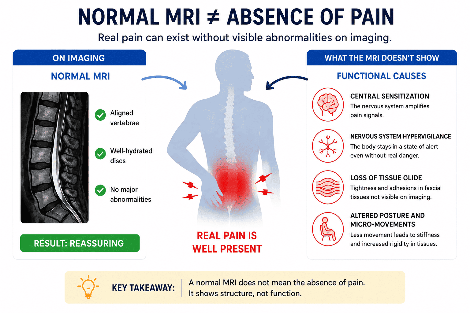
In other words, your back can look “normal” on the image while still generating functional pain: the structure may be intact, but the way it functions may be disturbed.
2. When your alarm system gets stuck on “ON”
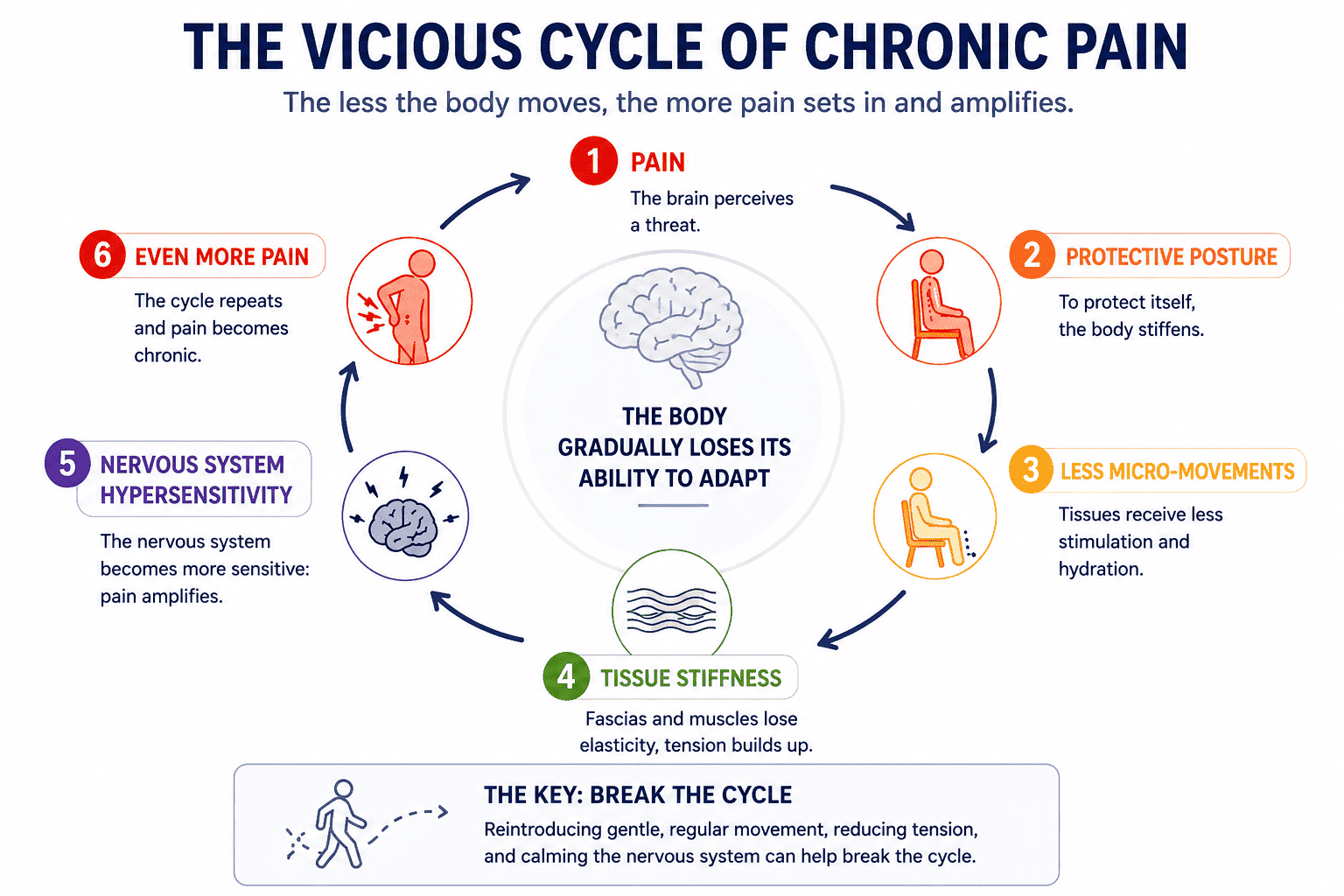
To understand why you still hurt despite normal exams, we need to look at the nervous system. Its role is to protect you. When it detects a threat, it triggers an alert: pain.
But after prolonged stress, repeated fixed posture, or an old pain experience, this system can become overly sensitive. This is known as central sensitization. The alarm threshold becomes lower, and the volume of the pain signal increases.
The brain may then interpret normal signals — such as the simple pressure of sitting — as a threat. Pain is no longer only the sign of an injury: it becomes an excessive response from a protective system that has become too vigilant.
This is one of the mechanisms through which pain can become chronic: through plasticity, the body “learns” to hurt. Pain circuits keep looping, like a computer program that no longer knows how to stop, even when the initial cause has disappeared.
3. Invisible tensions that “suffocate” your tissues
Another common cause of invisible pain lies in the fascia. Fascia is connective tissue that surrounds and links muscles, nerves, blood vessels, and organs. When healthy, it remains flexible and allows tissues to glide freely over one another.
The problem is that these tissues do not tolerate prolonged immobility well. When you remain seated for hours in a fixed position, water is progressively squeezed out of the tissues. They become denser, lose fluidity, and may feel more “sticky.”
This loss of tissue glide creates tensions that are invisible on MRI but very real for your pain receptors. Stiffer tissues may eventually compress or irritate small nerves and blood vessels, creating sensations of burning, deep pressure, or diffuse discomfort.
Your body can then turn into a kind of internal armor: less mobile, more reactive, and more sensitive.
4. Breaking the cycle through gentle mobility
When medical imaging is normal, the solution is usually not complete rest. The real challenge is to restore tissue fluidity and gradually reassure the nervous system.
This is where micro-movement plays an essential role. It is not about intense exercise. It is about reintroducing small, regular adjustments, especially in the sitting position:
- gently tilting the pelvis;
- frequently changing support points;
- breathing deeply;
- avoiding prolonged fixed positions.
These small movements send safety signals to the brain. They help fascia recover glide, support local circulation, and allow the alarm system to gradually lower its volume.
This is the logic behind bioactive sitting technologies such as Aporia®. Instead of holding the body in a rigid posture, they accompany natural micro-oscillations to transform sitting time into a form of continuous mobility.
Conclusion
Having back pain with a normal MRI is not a dead end. It often means that the body has lost part of its adaptive capacity: a hypervigilant nervous system, less mobile tissues, and less fluid fascia.
Understanding these mechanisms already changes the way we look at pain. By restoring pelvic movement, tissue fluidity, and reassuring signals to the brain, it becomes possible to gradually break the vicious cycle of chronic pain.
Learn more
-
Aporia® bioactive seat cushions for functional rehabilitation
Discover how bioactive sitting can support pelvic mobility, micro-adjustments, and the body’s natural adaptation mechanisms. -
Fascia and pain: understanding tissue glide and invisible tensions
Explore the role of fascia, tissue glide, and hidden mechanical constraints in chronic pain. -
Pelvic mobility and micro-movements in sitting
Learn why small pelvic adjustments can be more important than large movements for reducing fixed constraints while sitting.
Sources & References
- Clifford Woolf — Groundbreaking research on central sensitization of the nervous system .
- Hélène Langevin — Studies on fascias, connective tissue, and chronic pain mechanisms .
- Sundblad et al. — Research on sedentary behavior, micro-movements, and pain related to prolonged sitting .
- David Butler & Lorimer Moseley — Neuroscientific approach to pain education, notably featuring the textbook Explain Pain (Second Edition) .