


1. Understanding the concept of a zone of mechanical vulnerability
ESTABLISHED DATA MECHANICAL INTERPRETATION SBNFA™ FRAMEWORK
The course of the pudendal nerve involves several changes of direction and several close relationships with pelvic structures. The main points of anatomical interest include the ischial spine region, the sacrospinous and sacrotuberous ligaments, the falciform process, and Alcock’s canal (pudendal canal) (Robert et al., 1998; Zapletal et al., 2024).
To review the full course and the branches involved, see Functional Anatomy and Neuromechanics of the Pudendal Nerve .
On this page, the expression zone of mechanical vulnerability is preferred to “lock” when there is no evidence of pathological compression. It refers to a region in which anatomical relationships may make the nerve sensitive to certain stresses, without presupposing the mechanism at work in any given patient.
The term mechanical lock is reserved for the SBNFA™ functional interpretation: a zone of vulnerability becomes a lock when the local capacity for gliding, strain, redistribution, or recovery is no longer sufficient to absorb the loads placed on it. The lock therefore describes a temporarily unfavorable relationship between a geometry, a load, and an adaptive capacity; it is neither synonymous with a fixed lesion nor equivalent to a demonstrated compression.
Anatomical mechanical interference may involve compression, traction, shear, or local irritation. These mechanisms may be intermittent or permanent, but their existence cannot be inferred from the location of the pain alone.
Posture dependence is an important clinical feature. Worsening in the seated position is one of the essential Nantes criteria, but it cannot be used to locate the precise site of involvement with certainty, nor to demonstrate ligamentous compression (Labat et al., 2008).
Zone of vulnerability = a potentially sensitive anatomical configuration. Mechanical lock = a functional state in which local adaptive capacities become insufficient in the face of stress. Entrapment or compression = a clinical mechanism that must be demonstrated, and not an automatic consequence of the two preceding notions.
2. The ischial spine region and ligamentous relationships
ESTABLISHED ANATOMY BIOMECHANICAL HYPOTHESIS
The pudendal nerve passes close to the ischial spine and the sacrospinous and sacrotuberous ligaments. This region is an important anatomical zone in descriptions of pudendal nerve entrapment (Robert et al., 1998).
In some situations, the relationships between the nerve, the ligaments, and the neighboring tissues may contribute to local irritation. The term ligamentous clamp, formed by the sacrotuberous and sacrospinous ligaments, may be used as a descriptive anatomical model (Shafik et al., 2007), but it should not be presented as an automatic diagnosis or as a mechanism demonstrated in all people suffering from pudendal pain.
In the clinical literature, the ligamentous clamp refers to the space where the pudendal nerve, after passing around the ischial spine and through the lesser sciatic foramen, runs between the sacrotuberous ligament, below, and the sacrospinous ligament, above, accompanied by the pudendal artery and veins. Ziouziou et al. (2013) describe this region as a frequent site of nerve irritation.
Sitting may aggravate symptoms in some patients. This aggravation may reflect the interaction of several factors: local pressure, pelvic orientation, duration of exposure, muscle tension, nerve sensitivity, and individual recovery capacity. It has not been established that posterior pelvic tilt systematically increases ligament tension or reduces the space available for the nerve in all individuals.
From a mechanical standpoint, a change in pelvic orientation or in the tension transmitted by neighboring tissues may nonetheless alter the relative geometry of this region and the way stresses are distributed within it. This proposition is consistent with anatomy, but its magnitude, direction, and clinical significance vary from person to person; it therefore does not justify establishing a single posture, notably posterior pelvic tilt, as a universal cause.
The ligamentous model should therefore be formulated as a biomechanical hypothesis that may explain certain clinical pictures, and not as universal causation.
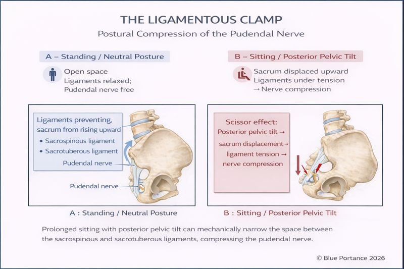
Worsening in the seated position remains an important clinical feature in the assessment of pudendal neuralgia. It is one of the Nantes criteria, along with pain located in the pudendal territory, the usual absence of nocturnal awakening, the absence of objective sensory deficit, and improvement after an anesthetic pudendal nerve block (Labat et al., 2008).
3. Alcock’s canal: fascial pathway and potential zone of mechanical interference
ESTABLISHED ANATOMY MECHANICAL INTERFACE
After passing near the ischial spine, the pudendal nerve runs through the pudendal canal, also known as Alcock’s canal. This canal corresponds to a splitting of the fascia associated with the obturator internus muscle and contains the pudendal neurovascular bundle (Robert et al., 1998; Zapletal et al., 2024).
According to Ziouziou et al. (2013), the nerve may also be involved slightly more distally, in Alcock’s canal, located about 15 mm further along and about 16 mm long. At this level, the fat surrounding the neurovascular bundle gives way to fibrous tissue. The authors describe venous stasis that may promote an entrapment syndrome in the seated position, particularly when the canal is narrowed or compressed by the falciform process of the sacrotuberous ligament. These elements describe a possible mechanism, not a systematic course.
Alcock’s canal can be described as a constrained anatomical interface. It is not necessarily a pathological tunnel, but a space in which the relationships between the nerve, the vessels, the fascia, and the muscular structures may become clinically relevant in certain situations.
Anatomical variations, a scar, a local change in the tissues, or mechanical stress could theoretically influence this environment. However, the available data do not support the assertion that pelvic floor hypertonia systematically reduces the volume of the canal or causes compression of the nerve.
It is also useful to distinguish two levels of analysis. The first is anatomical: it concerns the presence of an abnormality or of a structural relationship that could cause mechanical interference. The second is functional: it concerns the nerve’s tolerance to variations in pressure, tension, and movement.
This functional tolerance presupposes relative mobility between the nerve, its sheaths, the vessels, and the obturator internus fascia. Alcock’s canal should therefore not be represented as a rigid conduit: it is a living tissue interface, bound to the neighboring structures yet capable, under physiological conditions, of accompanying their relative movements.
In the SBNFA™ interpretation, the lock does not necessarily correspond to a permanent reduction in the caliber of the canal. It may also refer to a situation in which repeated or concentrated stress loads this interface faster than it can redistribute or recover from it. This mechanism makes an intermittent expression, dependent on the duration of exposure, conceivable, without demonstrating that it explains the individual case.
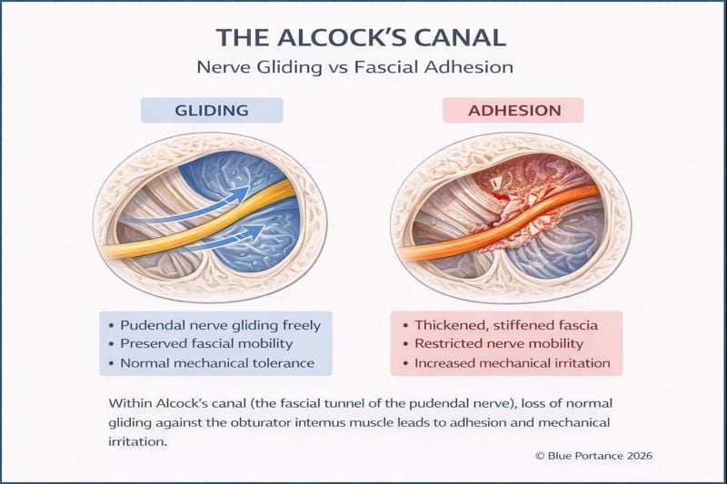
The fascial densification model may be invoked as a general hypothesis regarding possible changes in connective tissue interfaces. It should not, however, be presented as specific evidence of densification of Alcock’s canal in pudendal neuralgia (Pavan et al., 2014; Stecco et al., 2019).
The dynamics of this interface are developed in Fascia, Nerve Gliding, and Tissue Densification .
4. Cumulative stress and symptom variability
MULTIFACTORIAL MODEL SBNFA™ HYPOTHESIS
Anatomical factors should not be analyzed in isolation. A potentially vulnerable zone may be better tolerated in one person and less well tolerated in another, depending on nerve sensitivity, medical history, exposure to stress, recovery, and neuromuscular factors.
A clinical picture may therefore result from the interaction between an anatomical predisposition, repeated mechanical load, muscle tension, sensitization of the nervous system, and insufficient recovery. This formulation describes a multifactorial model and does not mean that several anatomical compressions are necessarily present.
Within SBNFA™, the “accumulation of locks” thus refers not to an automatic addition of lesions, but to the progressive reduction of compensatory possibilities. A first zone may remain silent as long as other segments or interfaces absorb and redistribute the load. When several possibilities for adjustment become limited, the same load may become more concentrated on the nerve pathway and be less well tolerated.
This interpretation connects four dimensions: geometry of the passages, relative mobility of the tissues, variability of stress, and recovery. It explains why sitting duration, repetition, the inability to vary support points, and the neuromuscular context can matter as much as the instantaneous intensity of a pressure.
Symptom variability — periods of remission, flare-ups in the seated position, the influence of exposure duration, and improvement with changes of position — may be compatible with an intermittent mechanical mechanism. It may also reflect factors of nerve sensitivity or pain modulation.
The Nantes criteria can help structure the clinical assessment, but they cannot determine the number of anatomical zones involved, nor distinguish with certainty between compression, irritation, sensitization, or a mixed mechanism (Labat et al., 2008).
The mechanical lock is a functional concept: it describes a local loss of adaptive possibilities, not necessarily an anatomical obstruction. In pudendal neuralgia, anatomical, mechanical, neuromuscular, and neurophysiological factors may interact, but their contribution must be assessed individually.
Scientific references
-
Robert R, Prat-Pradal D, Labat JJ, Bensignor M, Raoul S, Rebai R,
Leborgne J.
Anatomic basis of chronic perineal pain: role of the pudendal nerve.
Surgical and Radiologic Anatomy.
1998;20(2):93–98.
DOI:
10.1007/BF01628908
.
View PubMed record -
Labat JJ, Riant T, Robert R, Amarenco G, Lefaucheur JP, Rigaud J.
Diagnostic criteria for pudendal neuralgia by pudendal nerve entrapment
(Nantes criteria).
Neurourology and Urodynamics.
2008;27(4):306–310.
DOI:
10.1002/nau.20505
.
View PubMed record -
Zapletal J, Nanka O, Halaska MJ, Maxova K, Hajkova Hympanova L,
Krofta L, et al.
Anatomy of the pudendal nerve in clinically important areas:
a pictorial essay and narrative review.
Surgical and Radiologic Anatomy.
2024;46(2):211–222.
DOI:
10.1007/s00276-023-03285-7
.
View PubMed record - Pavan PG, Stecco A, Stern R, Stecco C. Painful connections: densification versus fibrosis of fascia. Current Pain and Headache Reports. 2014;18(8):441. DOI: 10.1007/s11916-014-0441-4 .
- Stecco A, Pirri C, Stecco C. Fascial entrapment neuropathy. Clinical Anatomy. 2019;32(7):883–890. DOI: 10.1002/ca.23388 .
-
Shafik A, El Sibai O, Shafik IA.
Role of sacral ligament clamp in the pudendal neuropathy (pudendal canal
syndrome): results of clamp release.
International Surgery.
2007;92(1):54–59.
View PubMed record -
Ziouziou I, Bennani H, Zizi M, Karmouni T, El Khader K, Koutani A,
Iben Attya Andaloussi A.
Le syndrome du canal d’Alcock ou névralgie pudendale : un diagnostic
à ne pas méconnaître [Alcock’s canal syndrome or pudendal neuralgia:
a diagnosis not to be overlooked].
Canadian Urological Association Journal.
2013;7(7-8):E486–E489.
DOI:
10.5489/cuaj.415
.
View PubMed record
The anatomical and clinical references above do not demonstrate that a “mechanical lock,” dynamic compression, or fascial densification explains every case of pudendal pain. They help distinguish established anatomical relationships from the proposed functional or biomechanical models.
The term “mechanical lock,” along with its integration into the SBNFA™ framework, constitutes a functional interpretive framework developed by Blue Portance. It does not correspond to a validated medical classification or to a stand-alone anatomical diagnosis.
Proposed internal reference:
- Blue Portance. SBNFA™ Framework — Neuro-anatomie, partie II : verrous mécaniques et contraintes pudendales [Neuroanatomy, Part II: Mechanical Locks and Pudendal Stresses]. 2026.
© Gil Ayache. The concepts, diagrams, terminology, and principles presented on this page constitute a work protected by copyright. They are made available to Blue Portance under an intellectual property license agreement, without transfer of economic rights or of authorship.
