


1. Distributed pain and referred pain
ESTABLISHED NEUROANATOMY FUNCTIONAL INTERPRETATION
The pudendal nerve carries sensory information from several regions of the perineum, the anal region, and the external genitalia. Irritation of the nerve or of one of its branches can therefore produce pain in a territory corresponding to this innervation.
It is nevertheless useful to distinguish between two situations. The first is distributed pain: the symptom is perceived in the peripheral territory of the irritated nerve. The second is referred pain in the stricter sense: the pain is felt in an area that does not correspond exactly to the site of stimulation or irritation.
In clinical practice, these two notions are sometimes used interchangeably. Yet pain located in the urethral, vulvar, penile, anal, or rectal region is not, on its own, enough to determine whether the pudendal nerve is involved, or to localize the level of any such involvement.
The distinction therefore does not cancel out the informative value of the territory. Distributed pain can link a distal perception to the branch that transmits the signal; referred pain or overlapping territories can make this relationship less direct. In both cases, the symptom map is a clue to be weighed against the other data, not topographic proof.
Symptoms must be weighed against how they change over time, their relationship to sitting, the clinical examination, any neurological signs, and possible local or visceral causes. The Nantes criteria can contribute to the assessment of pudendal neuralgia, but they do not replace differential reasoning (Labat et al., 2008).
The location of pain informs without localizing on its own: it provides information about the pain distribution and about the pathways likely to transmit the signal, but it is not enough to identify the structure responsible or the precise site of any nerve irritation.
2. The branches of the pudendal nerve
ESTABLISHED ANATOMY TEACHING MAP
The classic description of the pudendal nerve includes three main groups of branches: the inferior rectal nerve, the perineal nerve, and the dorsal nerve of the penis or clitoris. This presentation is useful for teaching purposes, but it must be qualified: the origin, course, and level of division of certain branches can vary between individuals (Robert et al., 1998; Zapletal et al., 2024).
The common course and the origin of these branches are detailed in Functional Anatomy and Neuromechanics of the Pudendal Nerve .
The trident therefore does not represent three completely separate lines. It schematizes three main terminal groups arising from a common course, with anatomical variations and overlaps. Its functional value lies precisely in this dual reading: distinguishing the dominant territories while looking for what may connect them upstream.
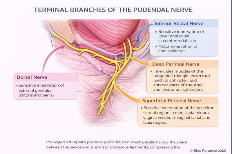
2.1 Inferior rectal nerve
The inferior rectal nerve generally crosses the ischioanal fossa to reach the anal region. It contributes to the motor innervation of the external anal sphincter and to the sensation of the lower part of the anal canal and the perianal region.
Its origin is variable: it can branch off from the pudendal nerve at different levels, before or within the pudendal canal. Anal or perianal pain may therefore be compatible with involvement of this branch, but it may also stem from a local anorectal or muscular cause, or from another nerve (Robert et al., 1998; Zapletal et al., 2024).
2.2 Perineal nerve
The perineal nerve has superficial and deep branches. It contributes to the sensory innervation of the skin of the perineum and of the scrotum or labia majora, as well as to the motor innervation of several perineal muscles and of the external urethral sphincter.
Perineal, vulvar, scrotal, or urethral pain may therefore lie within a territory compatible with this branch. However, a urinary or genital symptom should not be automatically attributed to the pudendal nerve: an infection, inflammation, a dermatological condition, myofascial pain, or another neuropathy must also be considered.
2.3 Dorsal nerve of the penis or clitoris
The dorsal nerve of the penis or clitoris is a predominantly sensory branch. It contributes to the sensation of the external genitalia and to the transmission of information related to touch, pressure, and sexual stimulation.
Hypersensitivity, allodynia, or pain on contact in the genital region may be compatible with pudendal involvement. It may also correspond to a local condition, vulvodynia, myofascial pain, or another nerve disorder (Aoun et al., 2021).
Sexual function results from the interaction of several somatic and autonomic pathways. It would therefore be incorrect to attribute the entire sexual response to the pudendal nerve alone (Aoun et al., 2021).
- A symptom that dominates in a single territory may direct attention toward a terminal branch, but does not demonstrate that the branch is involved.
- Symptoms affecting several pudendal territories and changing in a synchronized way may lead to examining a common mechanism located further upstream, or a broader functional interaction.
- Symptoms that are scattered, not synchronized, or extend well beyond the pudendal territories call for broadening the reasoning further to local, myofascial, or visceral causes, or to other nerve pathways.
These configurations are guides for reasoning. None of them constitutes a localization rule or a validated diagnostic criterion.
3. Clinical presentations and differential diagnoses
CONVERGING CLUES
Pain located in a pudendal territory can take very diverse forms: burning, electric-shock sensations, pain on pressure, hypersensitivity, numbness, or deep discomfort. Its neuropathic character must, however, be assessed clinically and cannot be inferred solely from the words the patient uses.
In an SBNFA™ reading, the analysis does not focus only on the location and quality of the pain. It also looks for the covariation of symptoms: do they appear together, do they increase after the same length of time sitting, do they decrease with the same changes in weight bearing, or do they behave independently? A shared pattern of change strengthens the plausibility of a shared mechanism, without determining its nature.
3.1 Urinary or urethral symptoms
Urethral burning or discomfort felt during or after urination may lie within a territory compatible with the perineal branches. However, a urinary tract infection, inflammation, mucosal irritation, bladder pain, or another urological cause must be looked for before suggesting an exclusively nerve-related origin.
3.2 Anal or rectal pain
Anal or rectal pain may be associated with the territory of the inferior rectal nerve. It may also correspond to a fissure, hemorrhoidal disease, inflammation, functional anorectal pain, a muscular disorder, or another local cause.
3.3 Genital pain
Vulvar, clitoral, penile, or scrotal pain may stem from irritation of pudendal branches, but also from a dermatological, infectious, inflammatory, or myofascial condition, or from another neuropathy. A normal local examination therefore does not, on its own, prove a pudendal origin.
A normal local examination does not mean that the pain is imaginary. It means that no obvious local cause was identified by the examination performed. The pain may then require a neurological, musculoskeletal, urological, gynecological, or proctological evaluation, depending on the clinical picture.
4. Why the diagnostic journey can be long
FUNCTIONAL SYNTHESIS
Perineal symptoms sit at the interface of several disciplines: urology, gynecology, proctology, dermatology, neurology, pain medicine, and pelvic floor rehabilitation.
This diversity can complicate the journey when each symptom is analyzed solely on the basis of the organ in which it is felt. Conversely, attributing symptoms too quickly to the pudendal nerve can also lead to overlooking a local cause or another neuropathy.
Two opposite forms of fragmentation are therefore possible: treating the urethra, the anus, and the genitalia separately even though their symptoms follow a common dynamic; or, conversely, grouping all pelvic and perineal pain too quickly under the pudendal label. The trident is useful when it allows clinicians to test the overall coherence without erasing the differential diagnoses specific to each territory.
Pudendal neuralgia is primarily a clinical diagnosis. There is no single test that can confirm every case, and several differential diagnoses must be considered before retaining this hypothesis (Labat et al., 2008; Fučík & Mašata, 2021).
The goal, therefore, is not to choose simplistically between an “organic” origin and a “nerve” origin. It involves relating the pain distribution, the time profile, sitting, the associated signs, the clinical examination, and the results of appropriate diagnostic testing.
A step-by-step reading can then be organized around four questions:
- Which territories are involved, and which local causes should be looked for?
- Do the symptoms in several territories change together?
- Does their onset depend on posture, the length of time sitting, weight bearing, or recovery?
- Does the picture correspond more closely to a terminal branch, to a more proximal common mechanism, to a mixed mechanism, or to another origin?
This sequence does not localize a lesion. It prevents the plurality of painful sites from being mistaken for a necessary plurality of causes, and it helps build a coherent hypothesis to be reassessed against the clinical data.
This approach also allows clinicians to acknowledge the reality of the pain when local examinations are reassuring, without asserting that a pudendal mechanism is necessarily present.
The possible consequences of a prolonged diagnostic journey are explored in The Diagnostic Odyssey and Central Sensitization in Pudendal Neuralgia .
Scientific references
-
Labat JJ, Riant T, Robert R, Amarenco G, Lefaucheur JP, Rigaud J.
Diagnostic criteria for pudendal neuralgia by pudendal nerve entrapment
(Nantes criteria).
Neurourology and Urodynamics.
2008;27(4):306–310.
DOI:
10.1002/nau.20505
.
View PubMed record -
Robert R, Prat-Pradal D, Labat JJ, Bensignor M, Raoul S, Rebai R,
Leborgne J.
Anatomic basis of chronic perineal pain: role of the pudendal nerve.
Surgical and Radiologic Anatomy.
1998;20(2):93–98.
DOI:
10.1007/BF01628908
.
View PubMed record -
Zapletal J, Nanka O, Halaska MJ, Maxova K, Hajkova Hympanova L,
Krofta L, et al.
Anatomy of the pudendal nerve in clinically important areas:
a pictorial essay and narrative review.
Surgical and Radiologic Anatomy.
2024;46(2):211–222.
DOI:
10.1007/s00276-023-03285-7
.
View PubMed record -
Aoun F, Alkassis M, Abi Tayeh G, Chebel JA, Semaan A, Sarkis J,
et al.
Sexual dysfunction due to pudendal neuralgia:
a systematic review.
Translational Andrology and Urology.
2021;10(6):2500–2511.
DOI:
10.21037/tau-21-13
.
View PubMed record -
Fučík T, Mašata J.
Pelvic neuropathic pain (differential diagnosis).
Česká gynekologie.
2021;86(4):279–283. DOI: 10.48095/cccg2021279.
View PubMed record
These references document the anatomy of the pudendal nerve, its branches, the clinical criteria for pudendal neuralgia, and the need for a differential diagnosis. They do not demonstrate that all urinary, rectal, or genital pain without a local lesion is necessarily of pudendal origin.
The SBNFA™ framework can be used as a functional interpretive framework for the relationship between the pain distribution, the nerve pathway, and the diagnostic journey. It does not constitute a validated medical classification.
Proposed internal reference:
- Blue Portance. Modèle SBNFA™ — Neuro-anatomie, partie III : douleur distribuée, douleur projetée et parcours diagnostique [SBNFA™ Framework — Neuroanatomy, Part III: Distributed Pain, Referred Pain, and the Diagnostic Journey]. 2026.
© Gil Ayache. The concepts, diagrams, terminology, and principles presented on this page constitute a work protected by copyright. They are made available to Blue Portance under an intellectual property license agreement, without transfer of economic rights or of authorship.
