


1. Fascia as a functional mechanical medium
TISSUE MECHANICS INTERFACES
The pelvic fascial system contributes to the mechanical organization of the relationships between neural, muscular, and bony structures. It is not merely a passive anatomical envelope. Its mechanical properties may influence the transmission, redistribution, and dissipation of stress between the different tissue planes.
From a clinical and functional standpoint, the relevance of fascia does not lie solely in its morphological appearance. It also lies in the possibility of relative movement between the tissues it separates or connects. This relative mobility may contribute to a nerve’s ability to adapt to movements of the pelvis and hip and to changes in posture.
The interface between the nerve and the surrounding tissues is therefore a relevant biomechanical element. It may be influenced by the organization of connective tissues, by local movements, by muscle tension, and by variations in pressure. However, the existence of a mechanical interface does not, on its own, justify concluding that a pathological entrapment is present.
Fascia thus fulfills a dual mechanical function: it provides a continuity that allows forces to be transmitted, while maintaining planes of relative mobility that prevent each movement from being transmitted in full to the nerve. Continuity and gliding are not in opposition; their combination allows stress to be redistributed and dissipated within a mobile tissue system.
The concept of fascial “entrapment neuropathy” describes the hypothesis that changes in the connective-tissue microenvironment surrounding a nerve may reduce its mechanical independence from adjacent tissues. This concept is general and does not constitute a specific demonstration concerning the pudendal nerve or Alcock’s canal (Stecco, Pirri & Stecco, 2019).
2. Neurodynamics: gliding as a functional property
PHYSIOLOGICAL PROPERTY MECHANICAL READING
Nerve gliding refers to a nerve’s ability to change its position relative to the surrounding tissues during movement. This property is part of the mechanical continuity of the nervous system, which must adapt to the variations in length, path, and tension induced by body movements (Butler, 2000).
When relative mobility is sufficient, mechanical variations can be better distributed among the different segments of the nervous system and the adjacent interfaces. This capacity for adaptation does not mean that the nerve is free of stress. Rather, it means that stress can remain compatible with the mechanical tolerance of the tissue.
Gliding is therefore not an incidental movement that the nerve could simply lose without mechanical consequence. It contributes to the nerve’s ordinary adaptation to changes in posture. In the SBNFA™ framework, the margin of displacement and strain still available constitutes a mobility reserve: it allows a change in length or path to be distributed across several segments rather than concentrated on a single interface.
Conversely, reduced relative mobility may theoretically alter the distribution of stress. Ordinary actions such as sitting down, straightening up, or walking may then become less well tolerated in some people. This situation may locally increase tensile, shear, or compressive loading.
The functional consequence is not necessarily immobility of the nerve, but a reduction in mechanical options: less relative displacement, more local strain, or faster stress concentration. This formulation links gliding to tolerance without presuming the existence of an adhesion or a compression.
However, movement-dependent pain should not automatically be equated with a loss of nerve gliding. Such an interpretation must be weighed against the clinical examination, the symptom course, and the other factors that may alter the sensitivity of the nervous system.
3. Fascial densification and possible loss of relative mobility
DOCUMENTED MODEL PUDENDAL APPLICATION TO BE VALIDATED
3.1 Moving beyond the opposition between local pain and radiating pain
In pudendal neuralgia, the opposition between local pain and radiating pain is often insufficient to describe the diversity of clinical presentations. Symptoms may vary with posture, the duration of exposure to sitting, physical activity, and the repetition of mechanical load.
The Nantes criteria provide a reference clinical framework for guiding the diagnosis of pudendal neuralgia. The five essential criteria include pain in the anatomical territory of the pudendal nerve, worsening in the sitting position, no nighttime awakening due to pain, no objective sensory deficit, and improvement after an anesthetic pudendal nerve block (Labat et al., 2008).
These criteria are useful for characterizing a clinical syndrome. They do not, on their own, demonstrate fascial involvement, a loss of nerve gliding, or a permanent anatomical compression. They must therefore be distinguished from the biomechanical models proposed to explain certain variations in symptoms.
3.2 What is fascial densification?
In the model proposed by Pavan et al. (2014), densification refers to a change in the mechanical properties of loose connective tissue, notably its viscosity and its gliding capacity. It must be distinguished from fibrosis, which corresponds to a more structural change in the organization and quantity of collagen.
Densification should therefore not automatically be equated with a scar, an irreversible adhesion, or established fibrosis. It is a model of tissue change that may impair relative movement between planes. Its clinical significance and its reversibility may depend on the context, the duration of mechanical stress, inflammation, mechanical load, and individual factors (Pavan et al., 2014).
Changes in the extracellular matrix, in the viscosity of connective tissue, and in collagen organization may theoretically influence friction and gliding between tissue planes. However, there is no justification for presenting a universal, proven sequence that systematically runs from dehydration to densification and then to fibrosis.
Densification is therefore better understood as a change in mechanical regime than as a mandatory step toward an irreversible lesion. It can serve as a hypothesis when mobility between planes appears less available, while remaining distinct from fibrosis and without being assertable in a given patient on the basis of symptoms alone.
3.3 Possible consequences for the pudendal nerve
A change in fascial interfaces may influence the mechanical environment of the neural structures that pass through or run alongside them. When relative mobility decreases, postural variations may be less well distributed among the tissues. A local increase in friction, shear, or tension then becomes theoretically possible.
The pudendal nerve runs through an anatomically complex region, in relation to the pelvic ligaments, the muscles, and the obturator internus fascia. This anatomical proximity may contribute to mechanical vulnerability in some patients, but it is not sufficient to demonstrate fascial involvement in an individual case (Robert et al., 1998).
To place these interfaces within the nerve’s full course, see Functional Anatomy and Neuromechanics of the Pudendal Nerve , as well as Mechanical Vulnerability and Pudendal Nerve Entrapment Mechanisms .
Neuropathic pain may therefore be compatible with a mechanical disturbance without a visible lesion. It may also result from other peripheral or central mechanisms. It is therefore preferable to speak of a possible loss of mechanical compatibility between the nerve and its environment rather than asserting that densification is the sole cause of the pain.
Possible change in interface properties → less available relative mobility → more limited stress redistribution → local loading potentially less well tolerated. The first three relationships constitute a plausible mechanical model; their presence along the pudendal course and their contribution to symptoms remain to be established on an individual basis.
4. Alcock’s canal: anatomical course and zone of vulnerability
ESTABLISHED ANATOMY DYNAMIC INTERFACE
4.1 An anatomical structure, not necessarily a pathological tunnel
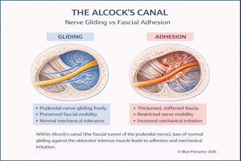
Alcock’s canal, also called the pudendal canal, is a passage formed by a splitting of the obturator internus fascia. The pudendal nerve and its vessels run through it in the lateral region of the pelvic wall.
The anatomical work of Robert et al. identified several potential entrapment sites along the course of the pudendal nerve, notably at the pudendal canal and the falciform process. These observations establish an anatomical basis for understanding certain forms of entrapment, but they do not mean that the canal is a zone of pathological compression in all patients (Robert et al., 1998).
A strictly anatomical reading, which would equate every instance of pudendal pain with permanent pinching in a rigid tunnel, is therefore insufficient. Symptoms also depend on the sensitivity of the nervous system, exposure to mechanical stress, posture, and the neuromuscular context.
4.2 An interface that may adapt to movement
The pudendal canal can be viewed as an anatomical interface within which the nerve must adapt to the movements and tension variations of the surrounding tissues. This adaptation probably involves several mechanical degrees of freedom, but the available data do not allow the canal to be described as a protective structure demonstrated under all conditions.
It nonetheless remains mechanically relevant not to represent it as an inert tunnel. The canal guides a neurovascular bundle within an environment linked to the obturator internus muscle: the nerve, the vessels, the fascia, and the neighboring tissues must therefore retain compatibility of movement. The term dynamic interface describes this functional requirement without attributing to the canal a specific protective function that has not been demonstrated.
Tissue densification, muscle hypertonia, a change in local pressure, or repeated postural loading could influence this interface. These mechanisms must, however, be formulated as functional hypotheses or possible contributing factors, not as established causality for all cases of pudendal neuralgia.
4.3 A posture-dependent vulnerability
The sitting position is one of the important clinical elements in the Nantes criteria, since worsening in the sitting position is one of the essential criteria for pudendal neuralgia (Labat et al., 2008). This clinical observation does not, however, demonstrate a single mechanism. It may reflect the interaction between pressure, pelvic posture, muscle tension, tissue load, and nerve sensitivity.
Pain may vary with the duration of exposure to a posture and with changes in position. These variations are compatible with a functional mechanical vulnerability, but they do not allow clinicians to locate the responsible structure with certainty, nor to distinguish compression, irritation, sensitization, or a combination of several mechanisms.
Alcock’s canal can thus be considered an anatomical point where several mechanical influences become clinically relevant. It is preferable to describe it as a potential zone of vulnerability rather than as an autonomous, systematic cause of pudendal neuralgia.
This approach also helps explain why the same anatomy may be well tolerated in one person and painful in another. The differences may involve nerve sensitivity, daily mechanical loads, tissue adaptive capacity, muscle coordination, and clinical history.
In SBNFA™, vulnerability appears when the stresses related to posture, their duration, and their repetition exceed this interface’s capacity for variation, gliding, and recovery. This is a model of loss of mechanical compatibility, not proof of densification or compression in Alcock’s canal.
5. Transition to chronic pain: interactions between pain, posture, and adaptation
MULTIFACTORIAL MODEL SBNFA™ HYPOTHESIS
5.1 A mechanical regime that may persist
Chronicity in pudendal neuralgia cannot be explained solely by progressive anatomical worsening. In some patients, the persistence of symptoms may reflect the interaction between repeated exposure to mechanical stress, nerve sensitivity, avoidance behaviors, sleep disturbances, muscle tension, and psychosocial factors.
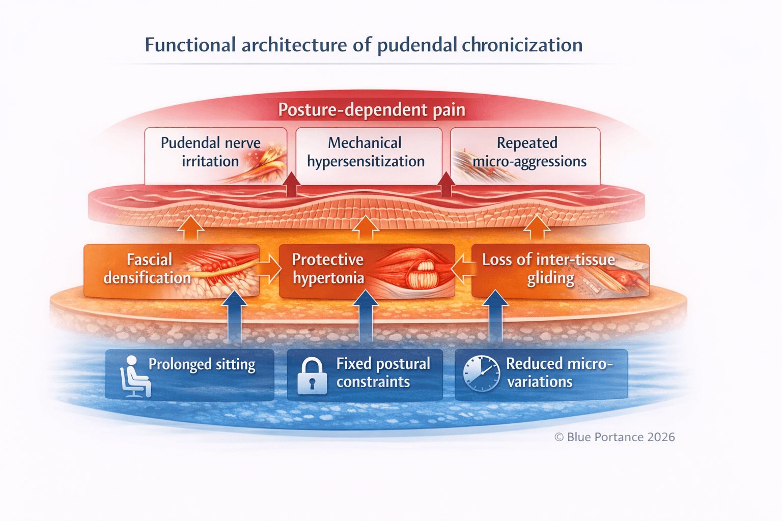
Prolonged sitting may represent a significant mechanical load when it reproduces or amplifies the symptoms. The duration of exposure, the workstation setup, the distribution of pressure, and the opportunity to change position may influence individual tolerance.
It has not been established, however, that a reduction in postural micro-variations systematically leads to fascial densification or fibrosis. This relationship should be presented as a biomechanical hypothesis to be investigated, not as a proven physiological law.
5.2 Possible feedback loops
Pain may induce a protective strategy and increased tension in certain muscles. This response may alter load distribution and reduce certain local movements. In turn, reduced activity or the repetition of protective postures may help maintain sensitivity and functional limitation.
This loop should not be understood as a simple, one-directional relationship. Pain can increase muscle tension, but muscle tension is not necessarily the initial cause of the pain. Likewise, the presence of muscle tension does not justify concluding that the pudendal nerve is compressed.
The fascial model proposes that a change in the connective-tissue microenvironment could reduce relative mobility between a nerve and the adjacent tissues. This model is relevant for generating clinical hypotheses, but it should not be presented as proof that pudendal pain stems from “adaptive fibrosis” in every case (Stecco, Pirri & Stecco, 2019; Pavan et al., 2014).
The loss of nerve mechanical tolerance in the pudendal nerve can therefore be viewed as the possible result of an imbalance between mechanical stress, the adaptive capacity of the tissues, and the sensitivity of the nervous system. This formulation helps integrate the biomechanical and neurophysiological dimensions without reducing chronicity to a single lesion.
The functional loop can be formulated without assuming a mandatory histological progression: pain → muscle guarding and reduction of available movements → lower variability of mechanical stress → more difficult recovery → lowered tolerance threshold → earlier onset of pain. Each of these relationships may vary from one person to another and be modulated by non-mechanical factors.
5.3 Why this reading changes the conceptual approach to management
This model helps explain why normal imaging does not rule out significant symptoms. Structural examinations may fail to identify certain functional changes, certain phenomena of nerve sensitivity, or certain posture-dependent interactions.
Conversely, normal imaging does not prove the existence of a fascial mechanism. Rather, it calls for interpreting the findings in relation to the clinical examination, the history of the symptoms, the provoking factors, and the responses to changes in load or posture.
A functional strategy may therefore seek to reduce aggravating mechanical stress, improve postural variability, progressively restore movement capacity, and take into account the sensitivity of the nervous system. These principles do not constitute a personalized therapeutic recommendation and must be adapted by qualified professionals.
Functional observation then focuses less on searching for “hidden fibrosis” than on how tolerance evolves: time before symptoms appear, number of positions still available, ability to vary support points, and time needed to recover. An improvement in these indicators suggests a greater functional margin; it does not demonstrate an anatomical change in the fascia.
Chronicity can thus be understood as the result of lasting interactions between mechanical, neuromuscular, and neurophysiological factors. This reading is more cautious than a model based on a single compression or on systematic fibrosis.
The complementary role of hypersensitivity is developed in The Diagnostic Odyssey and Central Sensitization in Pudendal Neuralgia .
Scientific References
- Butler DS. The Sensitive Nervous System. Adelaide: Noigroup Publications; 2000.
- Labat JJ, Riant T, Robert R, Amarenco G, Lefaucheur JP, Rigaud J. Diagnostic criteria for pudendal neuralgia by pudendal nerve entrapment (Nantes criteria). Neurourology and Urodynamics. 2008;27(4):306–310. doi: 10.1002/nau.20505 .
- Pavan PG, Stecco A, Stern R, Stecco C. Painful connections: densification versus fibrosis of fascia. Current Pain and Headache Reports. 2014;18(8):441. doi: 10.1007/s11916-014-0441-4 .
- Robert R, Prat-Pradal D, Labat JJ, Bensignor M, Raoul S, Rebai R, Leborgne J. Anatomic basis of chronic perineal pain: role of the pudendal nerve. Surgical and Radiologic Anatomy. 1998;20(2):93–98. doi: 10.1007/BF01628908 .
- Stecco A, Pirri C, Stecco C. Fascial entrapment neuropathy. Clinical Anatomy. 2019;32(7):883–890. doi: 10.1002/ca.23388 .
The references above are used to support general anatomical, clinical, or conceptual points. They do not demonstrate that fascial densification, fibrosis, or a loss of gliding is the cause of every case of pudendal neuralgia.
© Gil Ayache. The concepts, diagrams, terminology, and principles presented on this page constitute a work protected by copyright. They are made available to Blue Portance under an intellectual property license agreement, without transfer of economic rights or of authorship.
