


This article is part of the Expert Guide “Fascia.”
Chapter 9 — Blue Portance Knowledge BaseHow Can Fascia Become Altered? Inflammation, Densification, and Fibrosis
Summary — Several types of fascial alteration need to be distinguished. A viscoelastic change or a densification of loose connective tissue can affect gliding without amounting to fibrosis. An injury, for its part, can lead to a scar or an adhesion. Finally, matrix deposition and remodeling can become excessive and persistent when fibroblasts and myofibroblasts remain abnormally active. Fibrosis can then set in. These situations can sometimes interact, but they are not necessarily stages of a single progression.
1. Five concepts not to be confused
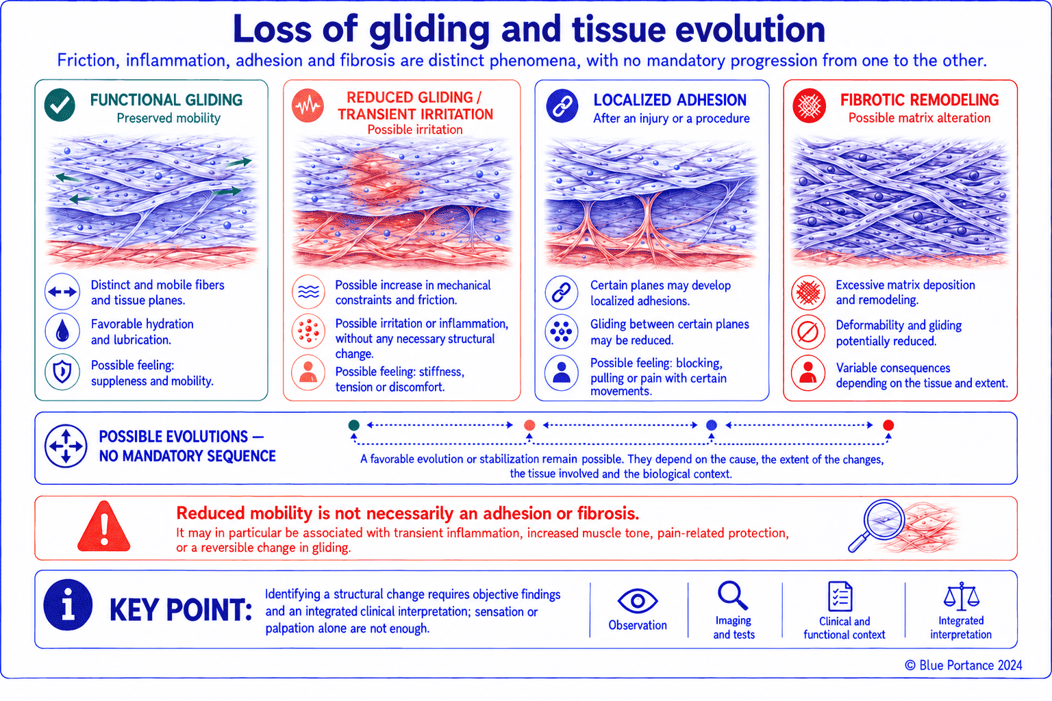
Reduced mobility can result from muscle guarding, edema, a change in viscosity, or pain. At this stage, it does not yet constitute an established, objectively demonstrable anatomical adhesion. A scar is the repair tissue formed after an injury. It can remain confined to the injured tissue, or contribute to an adhesion when it abnormally binds together two layers that normally move freely. Fibrosis, by contrast, is an excessive and persistent accumulation of matrix, which can disrupt the architecture and function of the tissue involved. This does not mean, however, that its collagen is necessarily disorganized in the microscopic sense of the term.
Densification, a separate concept
Densification deserves its own definition, because it lies at the heart of this chapter without being the same as the other four concepts. In the model proposed by Pavan and colleagues, it mainly refers to a change in the properties of the hyaluronan-rich loose connective tissue located between fascial layers. This change may increase its viscosity and impede gliding between those layers (Pavan et al., 2014).
This concept must be distinguished from collagen fibrosis. A later review describes the same phenomenon as an aggregation of hyaluronic acid. This aggregation can be observed in several organs, not only in fascia (Stecco et al., 2022). It is, however, a mechanistic model derived from narrative reviews and experimental observations, not a diagnostic nomenclature validated by consensus. The authors themselves acknowledge that there is no commonly accepted criterion. Clinically distinguishing densification from a simple physiological variation in viscosity in a given person therefore remains difficult.
Five concepts, five possible trajectories
These five concepts differ in their mechanism, their reversibility, and their permanence. Yet they are not the mandatory stages of a single trajectory. A person may have a scar without ever developing fibrosis, or tissue densification without an established adhesion. These developments depend on local and systemic factors specific to each situation.
| Concept | What is altered | Status |
|---|---|---|
| Functional change | Mechanical behavior, hydration state, muscle tone, pain | Potentially transient, reversible in the short term depending on the mechanism involved |
| Densification | Properties of the loose connective tissue and of hyaluronan between fascial layers | Mechanistic concept proposed in fascia research, without a universal clinical diagnostic criterion |
| Adhesion | Relative mobility between two surfaces that normally move in relation to each other | Anatomical abnormality or scar-related sequela; its course depends on its nature, extent, location, and tissue context |
| Scar | Repair tissue formed after an injury | Expected physiological response, which may remain functional or become pathological |
| Fibrosis | Excessive and persistent accumulation of extracellular matrix | Lasting structural change, with reversibility that varies depending on the tissue and the cause |
Confusing these states leads to overestimating the severity of simple, passing stiffness or, conversely, to underestimating tissue remodeling that is truly established. This confusion is common. Popular writing on fascia has sometimes fueled it, by too quickly equating any feeling of tension with an “adhesion” that needs to be treated (Lesondak, 2019).
2. The role of inflammation
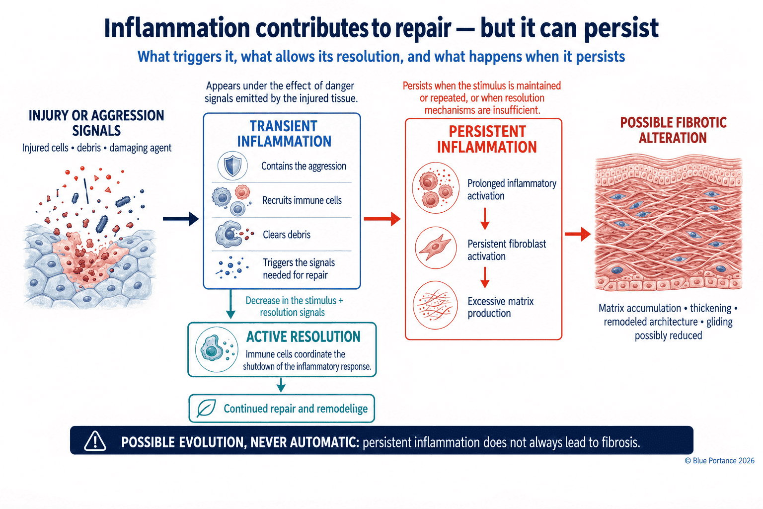
Mechanical loading of fascia does not systematically trigger a repair cascade. Depending on its intensity, it may simply produce a transient viscoelastic response or adaptive mechanotransduction, described in previous chapters, without any actual tissue injury occurring. A mechanical, thermal, chemical, or infectious insult must actually cause an injury for inflammation to come into play. Inflammation then recruits cells, increases vascular permeability, and organizes repair. At that point, it is necessary for healing. In its normal, time-limited course, it is an appropriate and beneficial biological response. It is therefore not a phenomenon to be systematically avoided or artificially suppressed. If it persists, cytokines and growth factors can promote excessive matrix production.
Four overlapping phases
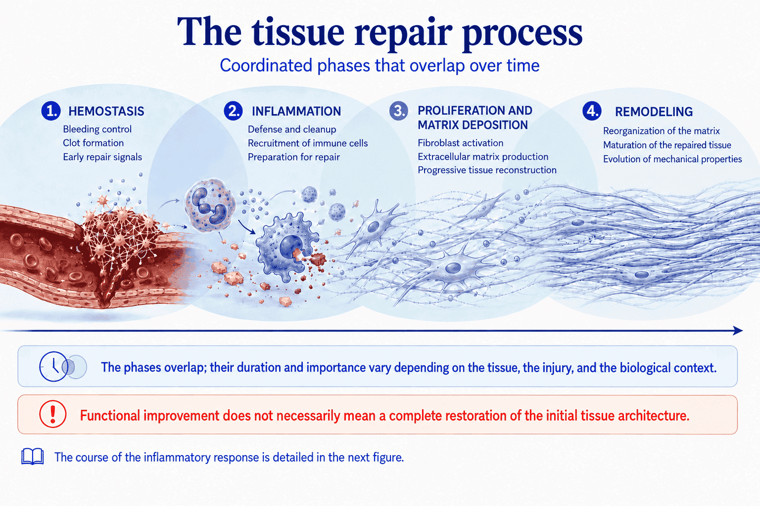
Tissue repair classically follows several phases that overlap in time. This model, described mainly in skin wound healing, provides a general framework through its main stages (Gurtner et al., 2008). Their duration, intensity, and cellular players nonetheless vary depending on the tissue, the type of injury, and its mechanical environment. A brief hemostasis phase occurs immediately after the injury. It is followed by an inflammatory phase, during which immune cells clear the injured area and release signals that recruit repair cells.
A proliferation phase follows, during which fibroblasts migrate to the area and begin producing new matrix. A longer remodeling phase then comes next. During this phase, the initial matrix, often disorganized, is gradually reorganized along functional lines of force. Under favorable conditions, this process results in a functional scar, strong enough and flexible enough not to hinder the normal function of the surrounding tissue.
When the inflammatory phase is prolonged
The inflammatory phase can be abnormally prolonged, due to infection, a general inflammatory background, or poorly suited mechanical loading of the area under repair. Remodeling can then remain prolonged or become pathological, with an imbalance between matrix production, organization, and degradation. Mechanical loading itself is not solely harmful. Its effect depends on its intensity, direction, frequency, and the stage of healing reached. Sufficiently graded loading can, on the contrary, favorably guide remodeling along functional lines of force. This unfavorable shift is generally not sudden. It builds up gradually, over weeks to months depending on the tissue, the extent of the injury, and the persistence of profibrotic signals. The tissue under repair continues to be loaded under conditions that do not allow it to complete its remodeling phase normally.
3. Fibroblasts and myofibroblasts
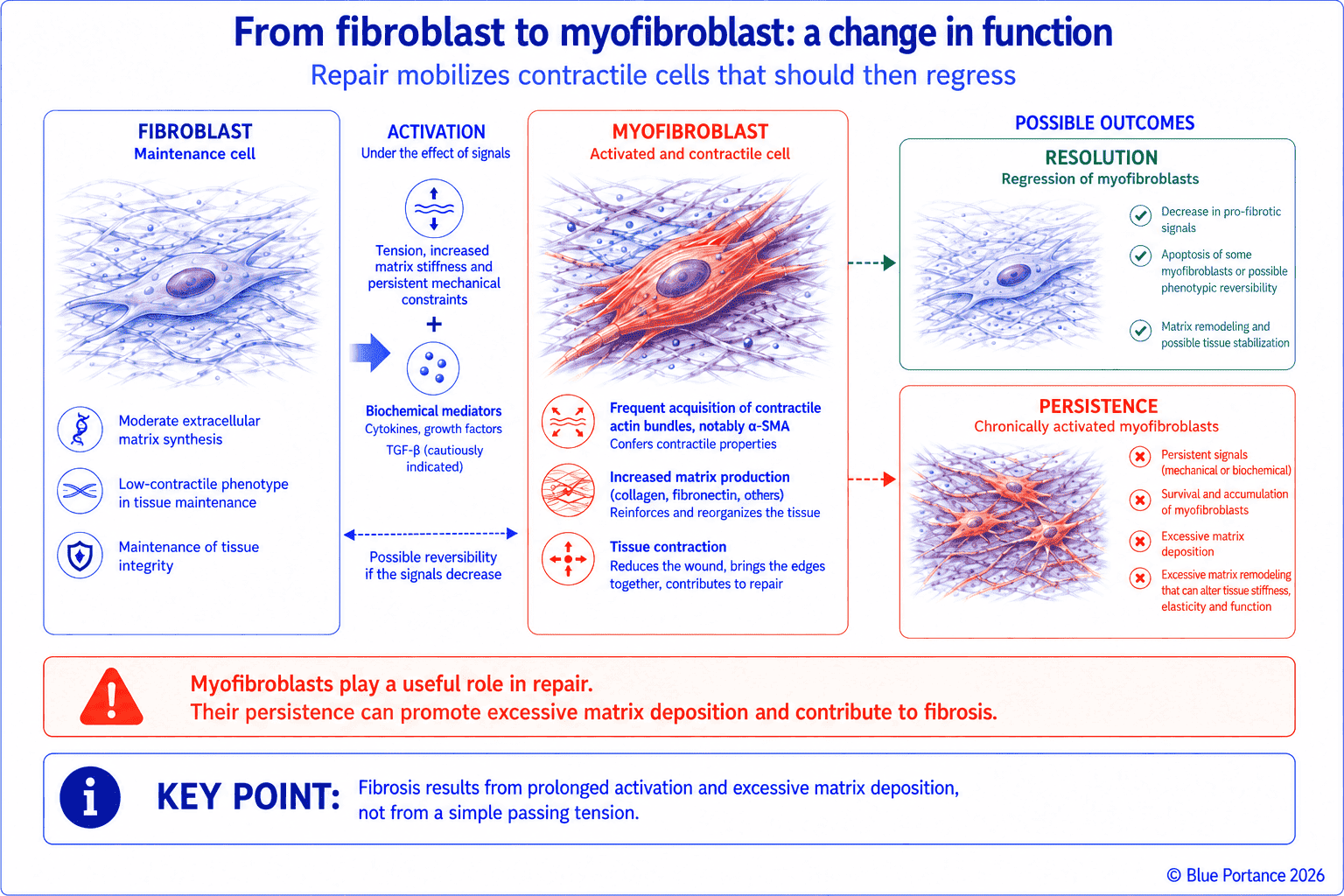
Under the combined effect of tension, stiffness, and mediators such as TGF-β, some fibroblasts acquire a contractile phenotype. Myofibroblasts actively draw the edges of a wound together thanks to their contractile apparatus, which is related to that of smooth muscle cells. However, if they persist abnormally beyond the healing phase, they can contract and stiffen the tissue in a lasting way (Tomasek et al., 2002). These cells are not specific to wound healing. A study combining histology and mechanography looked for myofibroblasts in human and animal fascia. It included 31 human donors and 20 animals, at several body sites. It then tested the contraction of isolated rat fascial tissue exposed to pharmacological stimulants. A positive correlation emerged between myofibroblast density and the measured contractile force (Schleip et al., 2019).
A study in humans and animals, but not everything measured in the same place
The exact scope of these results should be clarified, however. The presence of myofibroblasts was investigated in human and animal tissue. Contraction itself, on the other hand, was measured mechanographically only in isolated rat fascia. In humans, a contractile contribution therefore remains a plausible extrapolation from the observed cellular presence, not a directly measured force. This finding nonetheless has an important implication. It suggests that some fascial cells could contribute to slow, low-amplitude variations in tissue tension. This time scale is measured in tens of minutes to a few hours. It remains far removed from the speed of a voluntary muscle contraction.
A plausible implication, yet to be confirmed in humans
Their actual importance in perceived stiffness, pain, or human postural control remains to be established through direct measurements in humans. See also the chapter on mechanotransduction, which details the cellular relays through which mechanical stress can influence this phenotype change.
4. A self-sustaining loop
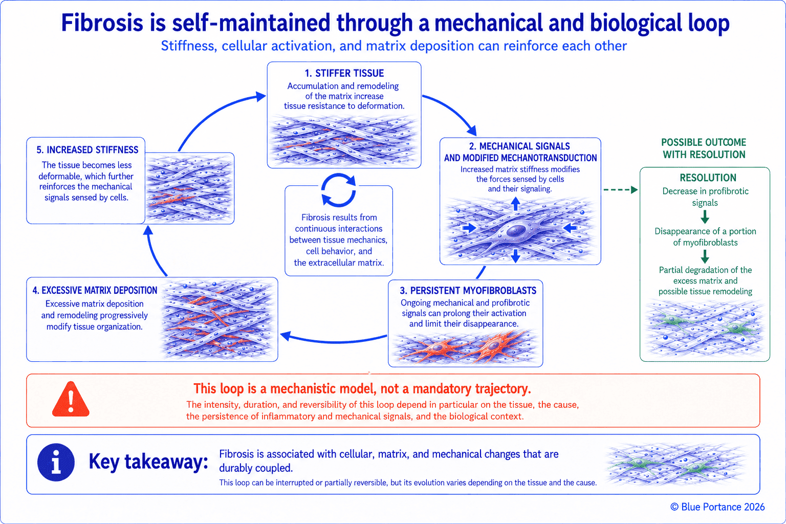
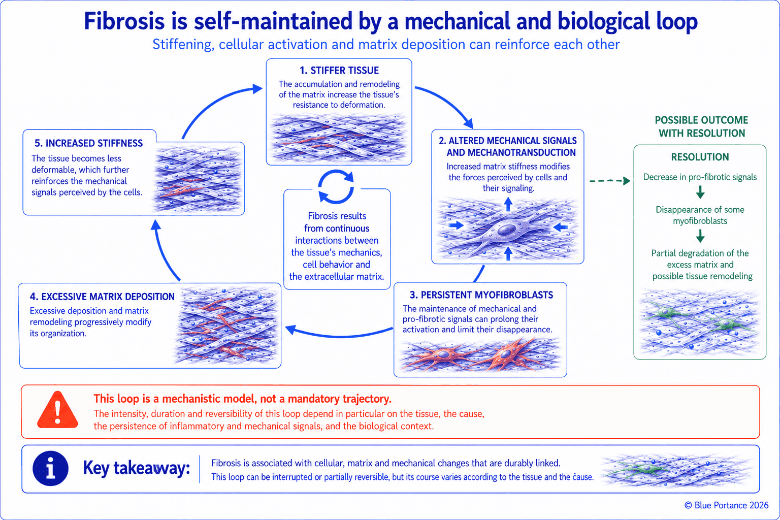
A stiffer matrix increases certain cellular tensions, which can in turn promote matrix production. This loop is one of the mechanisms that may sustain fibrosis that is already under way. It does not, however, demonstrate that a simple functional restriction will spontaneously progress to fibrosis.
This loop directly involves the mechanotransduction mechanism described in Chapter 6. A matrix made stiffer by an initial collagen deposit changes the forces sensed by the surrounding fibroblasts. It can thus reinforce certain mechanotransduction pathways that favor maintenance of the myofibroblastic phenotype (Chiquet et al., 2009). This can in turn contribute to further stiffening of the matrix. The fibrotic matrix itself thus helps maintain a profibrotic cellular phenotype, rather than being merely its consequence (Herrera et al., 2018).
In addition to this local loop, there is a plausible but less firmly established neighboring effect. A local reduction in mobility can alter the distribution of strains in neighboring tissues, described in Chapter 4. The extent and clinical relevance of this redistribution depend, however, on the region, the movement, and the adaptive capacity of the other structures. No data in the article, however, supports the claim that an initially circumscribed restriction necessarily affects, over time, a larger anatomical region.
5. What can and cannot be restored
A recent functional change can evolve with healing, a gradual return to movement, and a reduction in inflammation. Established fibrosis does not simply disappear through micro-movements. The goal then often becomes improving the function of the remaining tissues and overall tolerance of daily activity.
This distinction echoes the one drawn in Chapter 6 between immediate mechanical response and delayed biological remodeling, applied here to the question of recovery. A recent functional dysfunction, without established excessive matrix deposition, can improve with a gradual return of mobility and resolution of the underlying inflammation. Established fibrosis, by contrast, corresponds to a matrix architecture that has already been remodeled. It is not reversed simply by reproducing the conditions that allowed a functional dysfunction to heal.
What influences the reversibility of fibrosis
The actual reversibility of fibrosis does not depend on a single factor or a single time axis. It varies according to the tissue involved, the initial cause, and the duration and persistence of the stimulus. The maturity and degree of cross-linking of the deposited collagen also play a role. The same goes for local vascularization and the balance between matrix metalloproteinases and their inhibitors. Finally, whether myofibroblasts disappear or persist over time also matters. Some forms of fibrosis can partially regress when the causal stimulus disappears. Others leave a lastingly altered architecture. It is therefore not possible to state a general prognosis that applies to all fascial fibrosis.
Adapting care rather than aiming for complete restoration
In this second case, care more realistically aims to optimize the function of the available tissues and overall tolerance of activity. A complete return to the previous tissue architecture cannot be assumed or considered a realistic goal in every situation. Functional improvement can thus be a more realistic objective than complete restoration of the original architecture. This does not mean, however, that the architecture has been fully restored. This distinction is not merely a theoretical nuance: it shapes realistic goals and avoids the frustration that often comes with expecting complete restoration. This chapter explains general mechanisms. It does not prescribe any universal course of action. A return to movement or progressive loading should be adapted to the context — particularly after surgery, an infection, an acute injury, or a fibrosing disease — rather than applied uniformly.
Key Takeaways
- Loss of gliding, adhesion, scar, and fibrosis are different.
- Inflammation is useful for repair but can become profibrotic.
- Myofibroblasts contract and remodel the matrix.
- Reversibility depends on the stage and nature of the alteration.
Frequently Asked Questions
Does stiffness mean fibrosis?
Are densification and fibrosis the same thing?
Does a loss of gliding necessarily progress to fibrosis?
Can massage break up an adhesion?
Is fibrosis irreversible?
Scientific References
- Chiquet M, Gelman L, Lutz R, Maier S. From mechanotransduction to extracellular matrix gene expression. Biochim Biophys Acta. 2009;1793:911–920.
- Tomasek JJ, Gabbiani G, Hinz B, Chaponnier C, Brown RA. Myofibroblasts and mechano-regulation of connective tissue remodelling. Nat Rev Mol Cell Biol. 2002;3:349–363.
- Schleip R, Gabbiani G, Wilke J, et al. Fascia is able to actively contract and may thereby influence musculoskeletal dynamics: a histochemical and mechanographic investigation. Front Physiol. 2019;10:336.
- Lesondak D. Le Fascia. [Fascia.] Éditions Ressources Primordiales; 2019.
- Gurtner GC, Werner S, Barrandon Y, Longaker MT. Wound repair and regeneration. Nature. 2008;453(7193):314–321.
- Pavan PG, Stecco A, Stern R, Stecco C. Painful connections: densification versus fibrosis of fascia. Curr Pain Headache Rep. 2014;18(8):441.
- Stecco A, Cowman M, Pirri N, Raghavan P, Pirri C. Densification: hyaluronan aggregation in different human organs. Bioengineering (Basel). 2022;9(4):159.
- Herrera J, Henke CA, Bitterman PB. Extracellular matrix as a driver of progressive fibrosis. J Clin Invest. 2018;128(1):45–53.
← Previous chapter: How Can Fascia Contribute to Pain?
Back to the Fascia guide
Next chapter: What Role Does Fascia Play in the Pelvis and Pelvic Floor? →
Note: this content explains general mechanisms. It does not constitute a medical diagnosis or a therapeutic recommendation.
© Gil Ayache. The original concepts, biomechanical models, diagrams, terminology, graphic representations, foundational figures, original texts, and principles presented on this page are works protected by copyright. They are made available to Blue Portance under an intellectual property license agreement, without transfer of economic rights or of authorship.
