


This article is part of the Expert Guide “Fascia”.
Chapter 10 — Blue Portance Knowledge BaseWhat Role Does Fascia Play in the Pelvis and Pelvic Floor?
Summary — The pelvis brings together organs, muscles, ligaments, nerves, and vessels in a confined space. The parietal pelvic fascia, the endopelvic fascia, the pelvic diaphragm, and the perineal membrane organize this support across several tissue levels. Together, they allow the mobility needed for filling, continence, defecation, and sexual function. They also structure the relationships with the obturator internus, the levator ani, the pudendal nerve, and the coccyx.
1. An Anatomical Crossroads
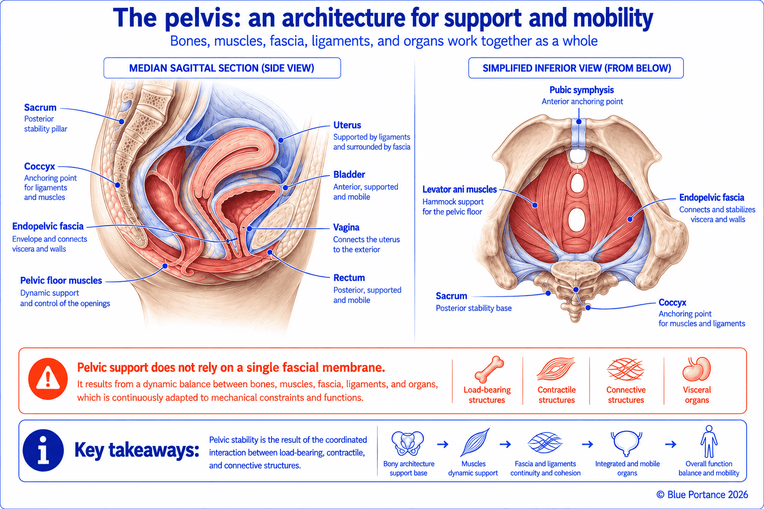
The pelvis must reconcile several demands that partly contradict one another. It must close off the abdomen from below while letting the urethra, vagina, or rectum pass through. It must also support mobile organs and accommodate large variations in abdominal pressure. This organization therefore does not rely on a single structure, but on several superimposed tissue levels.
The bony framework — pelvis, sacrum, and coccyx — thus forms the load-bearing structure of the whole. The parietal pelvic fascia, for its part, lines the inner surface of the obturator internus and levator ani muscles. The endopelvic fascia, often described as “visceral,” envelops the organs. It also connects them to one another and to the pelvic walls. The pelvic diaphragm, formed by the levator ani and the ischiococcygeus (coccygeus), closes off most of the pelvic cavity. The perineal membrane completes this closure lower down, between the ischiopubic rami. Finally, between the organs and the walls, subperitoneal spaces provide passage for vessels and nerves.
This organization is nonetheless less uniform than it appears. A recent systematic review points out that the description of the endopelvic fascia remains uncertain and varies from author to author (Roch et al., 2021). This variability concerns both its microanatomy and its macroscopic continuity. The term “endopelvic fascia” therefore refers more to a functional set of connective tissues than to a continuous, uniform membrane. This partly explains why anatomists, surgeons, and rehabilitation specialists do not always describe it in the same way (Ashton-Miller & DeLancey, 2007).
2. Supporting Mobile Organs
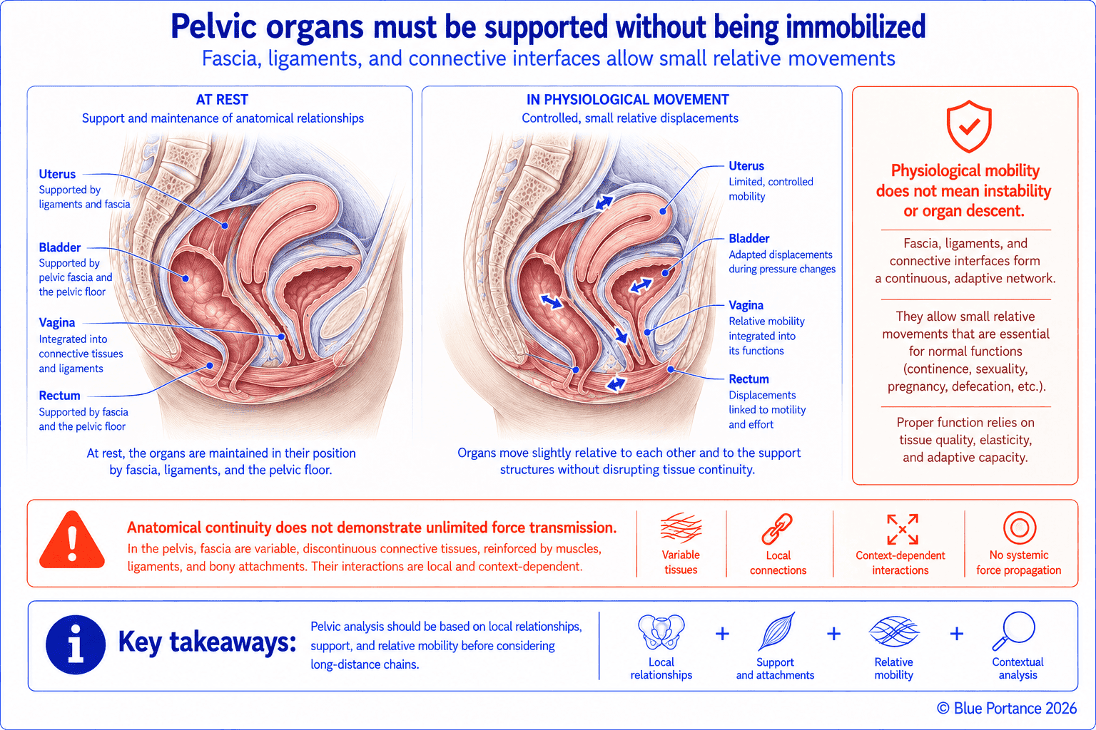
The bladder, rectum, and genital organs constantly change in volume and position. Their support is therefore not a rigid suspension. Connective attachments, the vaginal or prostatic walls, and ligaments work together with abdominal pressures and muscle activity. Together, they maintain the position of the organs without immobilizing them.
Support Shared Among Several Structures
The levator ani provides an active muscular floor that continuously counteracts increases in intra-abdominal pressure. Around it, connective attachments link the organs to the walls. In women, the uterosacral and cardinal ligaments contribute in particular to apical support (Herschorn, 2004; DeLancey, 1993). In men, the relationships between the bladder, prostate, and rectum are organized by other connective structures, including the puboprostatic ligaments, the periprostatic fascia, and the rectoprostatic fascia — although these arrangements are not strict anatomical equivalents (Raychaudhuri & Cahill, 2008). The perineal body, located between the vagina and the anus, or in an equivalent position in men, also serves as a central anchoring point where several of these connective attachments converge.
This distribution of roles also explains an important phenomenon. A single isolated injury is not always enough, on its own, to cause a support disorder. A muscle tear or a ligament injury, for example, often remains partially compensated for by neighboring structures (Ashton-Miller & DeLancey, 2007).
Supporting, Suspending, Allowing Movement, Accommodating Deformation
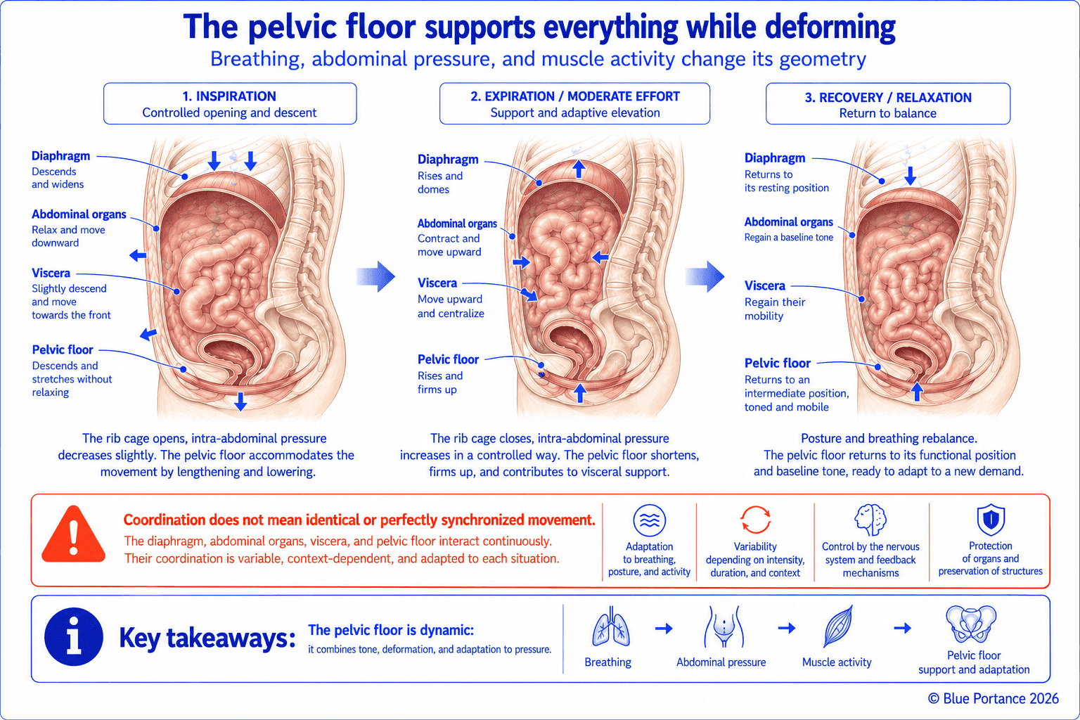
These four functions, however, are not the same thing. Supporting means bearing a weight without letting it descend. Suspending means holding an organ from above. This is achieved through ligaments rather than through support from below. Allowing movement means permitting a necessary physiological displacement, for example during bladder filling. Finally, accommodating deformation describes the ability of the pelvic floor to deform temporarily. This deformation occurs under the effect of breathing, exertion, or coughing, before a return to the resting position.
These four roles are therefore distributed among several structures rather than resting on a single tissue. Thus, the same abdominal load can be partially absorbed by muscle tone. It can also be transmitted to the ligaments or cushioned by the temporary deformation of the pelvic floor itself.
3. Interfaces with the Obturator Internus and Levator Ani
The obturator fascia lines the inner surface of the obturator internus muscle and thus contributes to the lateral wall of the pelvis. In particular, it provides an attachment zone for two tendinous arches that are often confused with each other: the arcus tendineus fasciae pelvis (ATFP) and the arcus tendineus levator ani (ATLA).
The arcus tendineus fasciae pelvis (ATFP) runs from the posterior surface of the pubic symphysis to the ischial spine. It thus serves as an attachment line for the pelvic fascia and the anterior vaginal wall. The arcus tendineus levator ani (ATLA) lies more medially and more posteriorly. It marks the line of origin of the levator ani muscle on the obturator internus. A three-dimensional imaging study confirmed that these two arches follow distinct paths. Their positions change in a characteristic way in the presence of a unilateral levator defect (Larson et al., 2012).
The Levator Ani and the Urogenital Hiatus
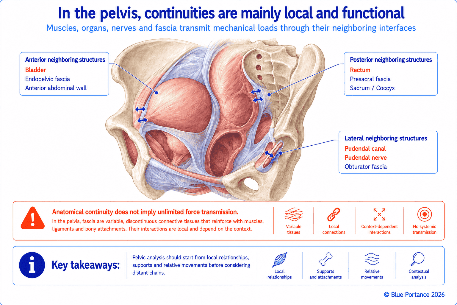
The levator ani, for its part, forms a dynamic muscular plane whose fibers insert in particular on the ATLA, the pubis, and the coccyx. Its fascial coverings extend toward neighboring tissues. Because of their neighboring attachments on the pelvic wall, mechanical changes in the obturator internus may thus, in theory, alter certain local interfaces with the pelvic floor — although this relationship alone cannot explain a symptom. Between the bundles of the levator ani, the levator hiatus provides the passages needed for the urogenital and anorectal tracts. Its anterior part, the urogenital hiatus, allows the urethra to pass, as well as the vagina in women. Further back, the anorectal junction crosses the levator plane. This zone is in fact a mechanically less resistant region.
This continuity, however, remains local. Stiffness of the hip or of the obturator internus is therefore not necessarily transmitted to all of the pelvic organs. Distance, fiber orientation, and several intermediate interfaces limit the propagation of stress (Roch et al., 2021).
4. The Pudendal Nerve and Its Fascial Interfaces
The pudendal nerve arises from the sacral plexus (S2–S4). It leaves the pelvis through the greater sciatic foramen, below the piriformis muscle. Its course then brings it close to the ischial spine and the sacrospinous ligament. It re-enters the pelvis through the lesser sciatic foramen, between the sacrospinous and sacrotuberous ligaments.
It then runs through the pudendal canal, or Alcock’s canal. This canal is formed within the fascia covering the medial surface of the obturator internus muscle. Its precise configuration is described differently from one study to another, but it corresponds to the course of the pudendal neurovascular bundle within the fascial environment of the obturator internus (Colebunders et al., 2011). This anatomy therefore requires the nerve to have both mobility and space. Any local reduction of this space may, in theory, interfere with its course.
Pain along the course of the pudendal nerve, however, does not on its own establish a neuropathy or an entrapment. On the contrary, the Nantes criteria require a combination of several clinical findings before this diagnosis is considered (Labat et al., 2008). In addition, other nerves, notably the inferior cluneal nerves, run through neighboring regions without sharing the same fascial course. They should therefore not be confused with the pudendal nerve.
5. The Coccyx at the Crossroads of Tensions
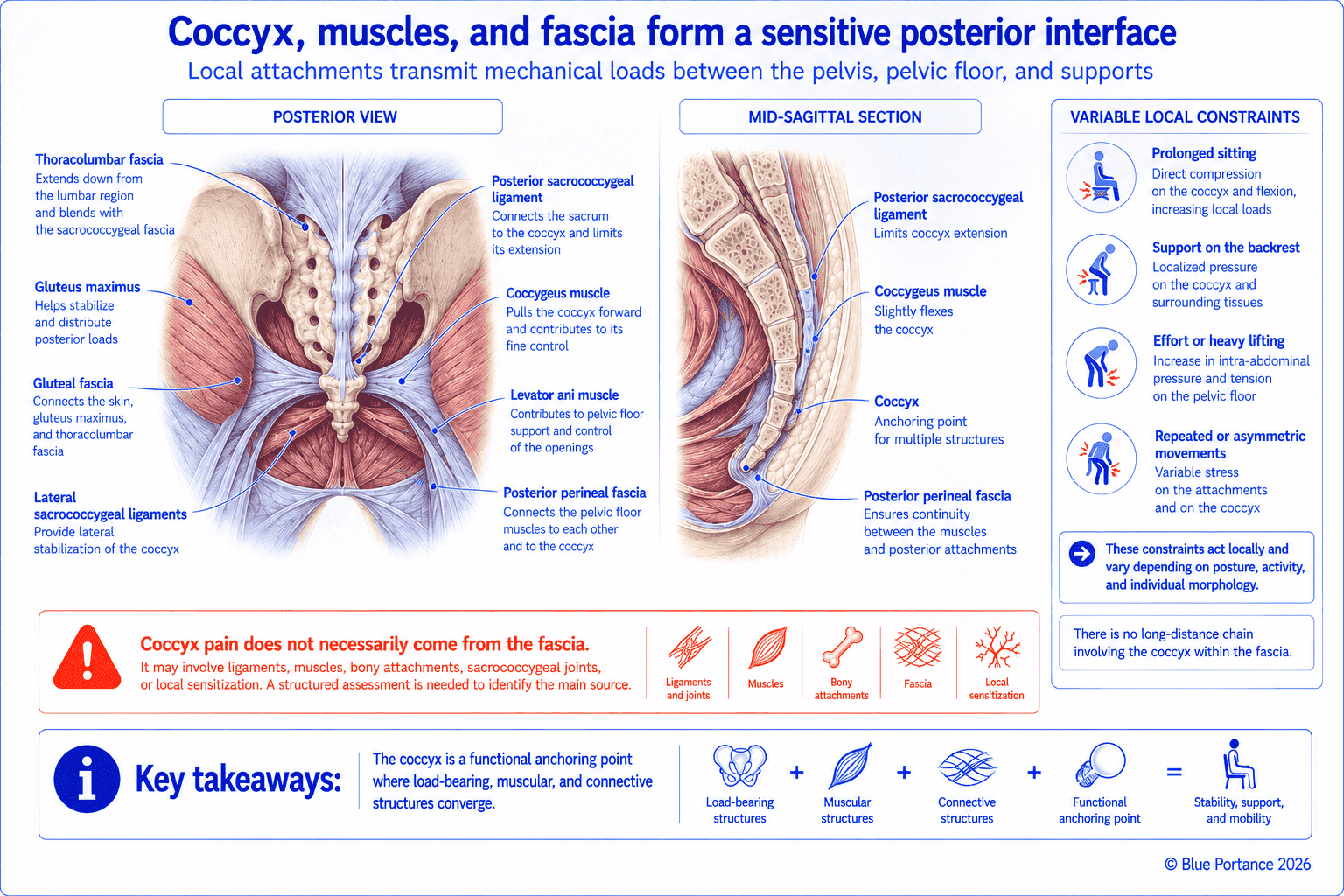
The coccyx extends the sacrum and receives numerous muscular, ligamentous, and fascial insertions. Together with the sacrum, it thus forms the posterior boundary of the pelvic cavity and an anchoring point where several structures converge.
Insertions Coming from Several Directions
Inserting on its anterior and lateral surfaces are, in particular, the coccygeus — or ischiococcygeus — muscle and some fibers of the levator ani. The coccyx thus receives attachments from several components of the pelvic diaphragm. The anococcygeal raphe also connects the external anal sphincter to its tip. Posteriorly, the gluteus maximus takes part of its origin from the lateral border of the coccyx. The sacrococcygeal ligaments — ventral, dorsal, and lateral — also stabilize the sacrococcygeal joint. Fibers of the sacrospinous ligament also extend to it (Woon & Stringer, 2012).
Limited but Real Mobility
The sacrococcygeal joint normally allows slight flexion-extension, particularly during defecation or sitting. Sitting loads, however, are not distributed evenly. When leaning backward, a larger share of body weight rests on the coccyx. Conversely, leaning forward generally reduces the coccyx’s contribution to support, while the ischial tuberosities remain the main load-bearing structures (Woon & Stringer, 2012). Pelvic posture, whether in anterior or posterior pelvic tilt, thus changes the mechanical environment of the coccyx. This does not, however, necessarily imply a disorder.
The coccyx should therefore not be described as a simple, isolated bony point. Its involvement in this crossroads of tensions does not mean that local discomfort is automatically explained by a fascial or postural cause. Several possible origins — articular, muscular, or traumatic — must be considered before drawing any conclusion.
Key Takeaways
- Pelvic support is dynamic and involves multiple tissues. It does not rest on a single fascial membrane, but on several superimposed tissue levels.
- Supporting, suspending, allowing movement, and accommodating deformation are thus four distinct functions, distributed among several structures.
- The ATFP and the ATLA are two distinct tendinous arches that are often confused. They structure the interfaces between the obturator internus and the levator ani.
- Alcock’s canal is formed within the obturator fascia. It remains an important anatomical interface, not automatic proof of pudendal nerve entrapment.
- The coccyx, which is mobile and has numerous anatomical attachments, should neither be viewed in isolation nor be singled out as the automatic cause of postural discomfort.
Frequently Asked Questions
Does fascia alone support the organs?
Does pelvic floor tension always come from fascia?
Do the ATFP and the ATLA refer to the same structure?
Is the coccyx part of the pelvic floor?
Scientific References
- DeLancey JOL. Anatomy and biomechanics of genital prolapse. Clin Obstet Gynecol. 1993;36:897–909.
- Ashton-Miller JA, DeLancey JOL. Functional anatomy of the female pelvic floor. Ann N Y Acad Sci. 2007;1101:266–296.
- Standring S, ed. Gray’s Anatomy: The Anatomical Basis of Clinical Practice. 42nd ed. Elsevier; 2020.
- Labat JJ, Riant T, Robert R, et al. Diagnostic criteria for pudendal neuralgia by pudendal nerve entrapment. Neurourol Urodyn. 2008;27:306–310.
- Herschorn S. Female pelvic floor anatomy: the pelvic floor, supporting structures, and pelvic organs. Rev Urol. 2004;6(Suppl 5):S2–S10.
- Roch M, Gaudreault N, Cyr MP, Venne G, Bureau NJ, Morin M. The female pelvic floor fascia anatomy: a systematic search and review. Life (Basel). 2021;11(9):900.
- Larson KA, Luo J, Yousuf A, Ashton-Miller JA, DeLancey JO. Measurement of the 3D geometry of the fascial arches in women with a unilateral levator defect and “architectural distortion”. Int Urogynecol J. 2012;23(1):57–63.
- Colebunders B, Matthew MK, Broer N, Persing JA, Dellon AL. Benjamin Alcock and the pudendal canal. J Reconstr Microsurg. 2011;27(6):349–354.
- Woon JTK, Stringer MD. Clinical anatomy of the coccyx: a systematic review. Clin Anat. 2012;25(2):158–167.
- Raychaudhuri B, Cahill D. Pelvic fasciae in urology. Ann R Coll Surg Engl. 2008;90(8):633–637.
← Previous chapter: How Can Fascia Become Altered? Inflammation, Densification, and Fibrosis
Back to the Fascia guide
Next chapter: How Does Sitting Load Fascia and Other Tissues? →
Note: this content explains general mechanisms. It does not constitute a medical diagnosis or a therapeutic recommendation.
© Gil Ayache. The original concepts, biomechanical models, diagrams, terminology, graphic representations, foundational figures, original texts, and principles presented on this page are works protected by copyright. They are made available to Blue Portance under an intellectual property license agreement, without transfer of economic rights or of authorship.
