


This article is part of the Expert Guide “Fascia.”
Chapter 4 — Blue Portance Knowledge BaseWhat Role Does Fascia Play in Tissue Dynamics and Protection?
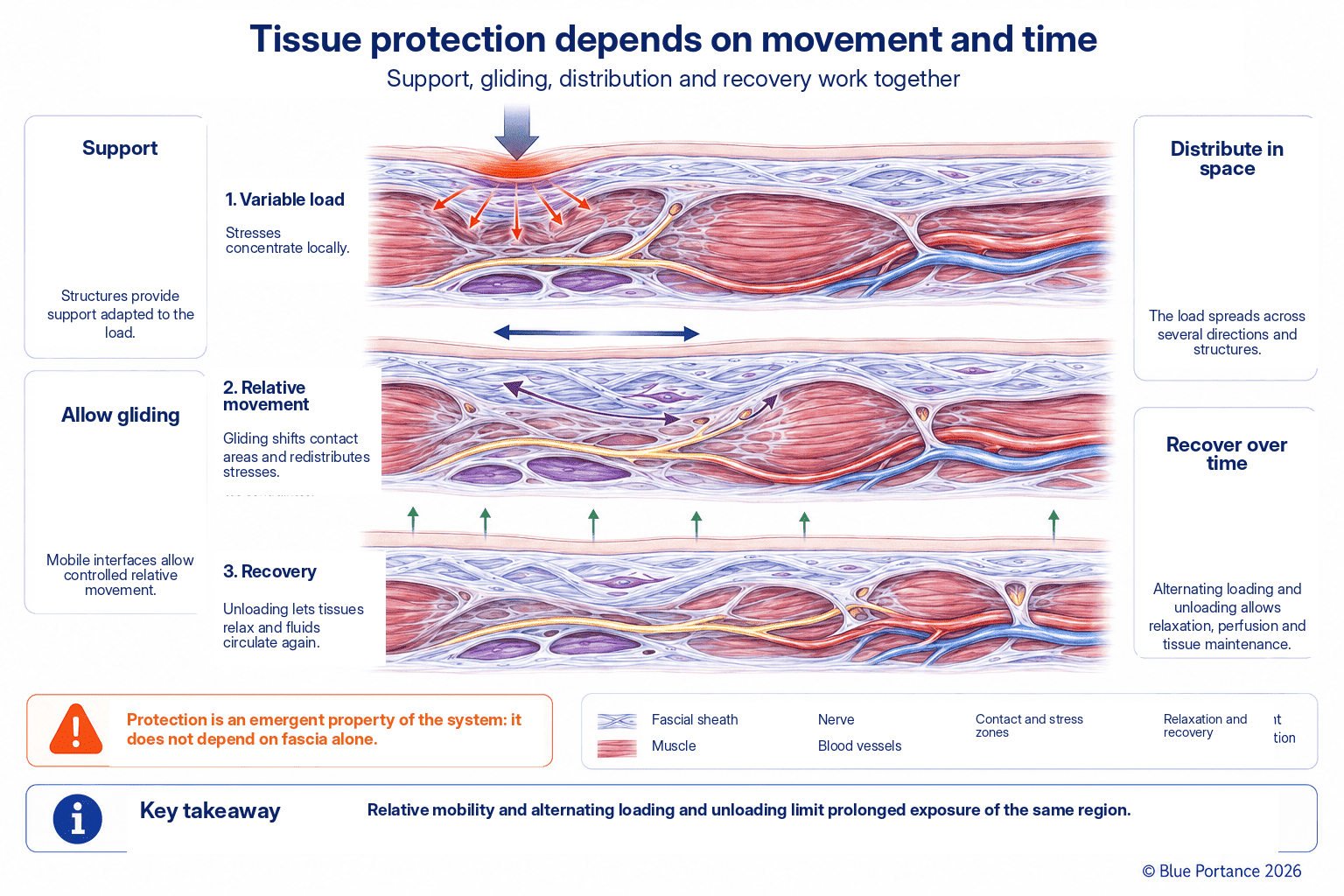
Summary — Organs, muscles, nerves, and vessels continuously change shape and position. Fascia maintains their anatomical relationships while allowing relative mobility. This combination of support, relative mobility, and redistribution can prevent certain displacements or certain stresses from concentrating on a single interface. In this way, together with the other tissues, it helps preserve sensitive structures.
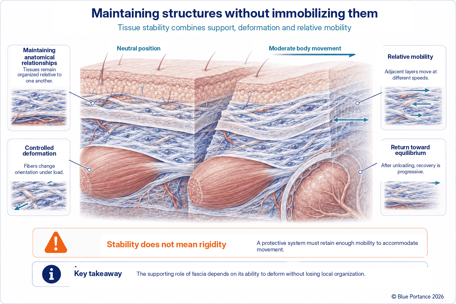
1. Holding Without Immobilizing
A living organization must, in fact, preserve form without fixing structures in place. Envelopes and septa thus delimit spaces, while looser layers allow displacement. Stability therefore results from a compromise between holding and mobility, a balance this chapter examines in turn at the level of tissue gliding, neurovascular accompaniment, and the limitation of friction.
This compromise is found at every scale. It applies to the broad definition of the fascial system adopted in this guide, which includes, in particular, fasciae in the strict sense, aponeuroses, retinacula, sheaths, and certain specialized connective envelopes (Adstrum et al., 2017). A joint capsule, for example, must keep the opposing bone surfaces aligned while tolerating the full range of joint motion. Likewise, a retinaculum must hold a tendon against the bone without preventing its longitudinal gliding during muscle contraction. In both cases, insufficient holding would compromise the stability of the structure. Conversely, excessive holding would restrict movement to the point of making it painful or ineffective. Their mechanical behavior is therefore functional only in relation to their role, their location, the direction of loads, and the range of motion required. There is thus no optimal level of stiffness in absolute terms.
The Consequences of an Imbalance
An imbalance in one direction or the other has distinct consequences. A tissue that has become too lax relative to its holding function — for example, after repeated distension — may allow excessive displacement between two structures that normally move together. The risk of local instability is therefore increased.
Stiffening and Loss of Relative Mobility
Conversely, a loss of relative mobility — for example, when a tissue becomes too stiff or too adherent — can alter local kinematics and redistribute strain toward other interfaces. The magnitude and extent of this redistribution, however, depend on the anatomical organization and the applied loads, and are neither automatic nor identical from one situation to another. This second scenario is taken up again in Chapter 9, which covers fibrosis and the loss of tissue gliding. Between these two extremes, most tissues function within a relatively broad range of acceptable resistance. This explains why substantial individual variability can coexist with normal function.
2. Allowing Internal Gliding
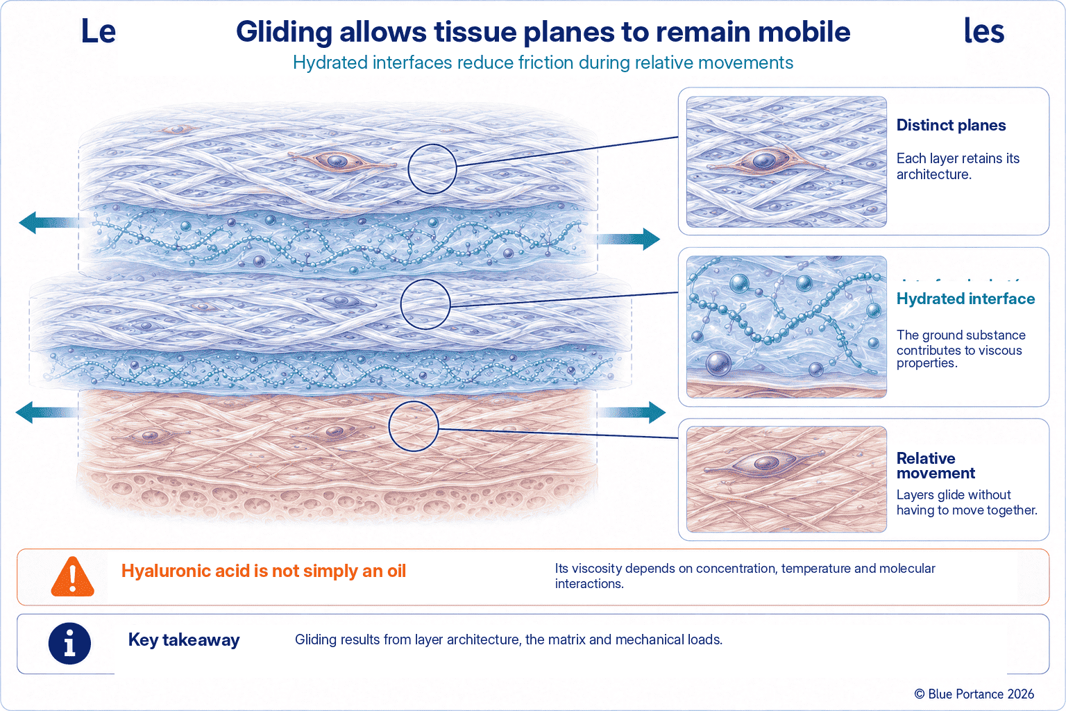
Beneath the skin, between muscles, or around an organ, layers move relative to one another. Gliding thus results from the coordinated deformation of fibers, ground substance, and fluids.
Direct endoscopic observations of loose connective tissue have described this mechanism. Performed intraoperatively in the gliding tissues of the hand, they show that between two fascial planes, a three-dimensional fibrillar network delimits spaces of variable geometry. These spaces are filled with hydrated ground substance. During movement, the fibers and the volumes they enclose continuously change shape and arrangement. This mechanism allows relative displacement while maintaining tissue continuity, rather than two smooth surfaces sliding over each other (Guimberteau et al., 2010). It is this continuous reorganization of the network, more than the mere presence of a lubricating film between two layers, that may help explain the range of gliding observed between certain tissue planes. Because these observations were made mainly in the gliding tissues of the hand, their generalization to all fascial interfaces in the body should be approached with caution.
When Gliding Is Lost
A loss of gliding increases the coupling between structures: the movement of one imposes more deformation on the other. Two tissues that normally move independently then behave as if they were partially bound together. The displacement of one then mechanically drags the other along, which can alter stress distribution well beyond the area initially involved. This increased coupling can thus alter mechanical transmission beyond the interface initially involved, more than an isolated local lesion would. On its own, however, it does not predict the extent or the functional consequences of this change for more distant structures.
3. Accompanying Nerves and Vessels
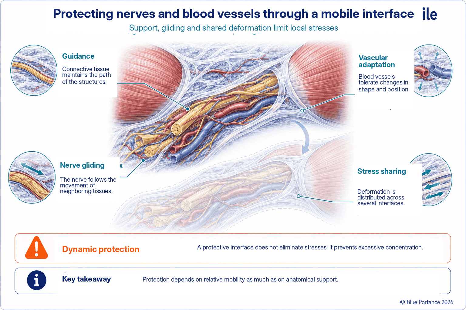
A nerve must be able to slide, change length slightly, and adapt to movements along its course. Vessels, for their part, must keep their lumen open despite changes in the shape of neighboring structures. The connective environment thus organizes these passages and limits focal stress.
The course of the median nerve at the wrist illustrates this requirement well. During wrist flexion and extension, the nerve glides several millimeters within the carpal tunnel to follow the movement of the neighboring flexor tendons. A total excursion of about 5.6 mm has been measured by ultrasound between wrist extension and flexion (Wang et al., 2014). Without this gliding, the nerve would be subjected to repeated stretching or compression.
This excursion depends on the mechanical properties of the nerve and its sheaths. It also depends on the nerve’s relative mobility with respect to the tendons and surrounding connective tissues. The pressure within the tunnel and the position of the proximal joints also come into play (Topp & Boyd, 2006). The same principle applies, at other scales and with other determinants, to most peripheral nerve pathways. Their connective environment must thus both hold them in place and allow this relative longitudinal gliding. It must also tolerate a degree of stretching, transverse deformation, and compression during normal movement.
How Vessels Adapt to Movement
Vessels impose a complementary requirement: adapting to the changes in length, curvature, and position induced by movement while maintaining perfusion compatible with the tissue’s needs. An artery crossing the back of the knee, for example, genuinely changes geometry between extension and flexion. This adaptation is not unlimited, however. In deep flexion, deformations, curvatures, and sometimes true kinks can appear along the femoropopliteal segment. These deformations are then accompanied by a local change in flow conditions (Nagita et al., 2024).
Prolonged Compressive Load While Sitting
The connective environment surrounding the vessel is thought to help distribute these changes in shape, although no direct biomechanical reference has yet precisely demonstrated this protective mechanism for the popliteal artery. In a region subjected to prolonged pressure, such as the ischial region while sitting, perfusion and nerve tolerance depend on several factors. These include, in particular, the intensity and duration of compression, the distribution of support, and the opportunities for offloading. The relative mobility of tissues may contribute to this adaptation, without being its sole determinant.
This protection has its limits, however. A narrow fibrous canal or prolonged pressure can, on the contrary, contribute to compression. This is particularly the case when the nerve pathway passes through a region where the available space is already reduced by the surrounding anatomy.
4. Limiting Friction and Shear
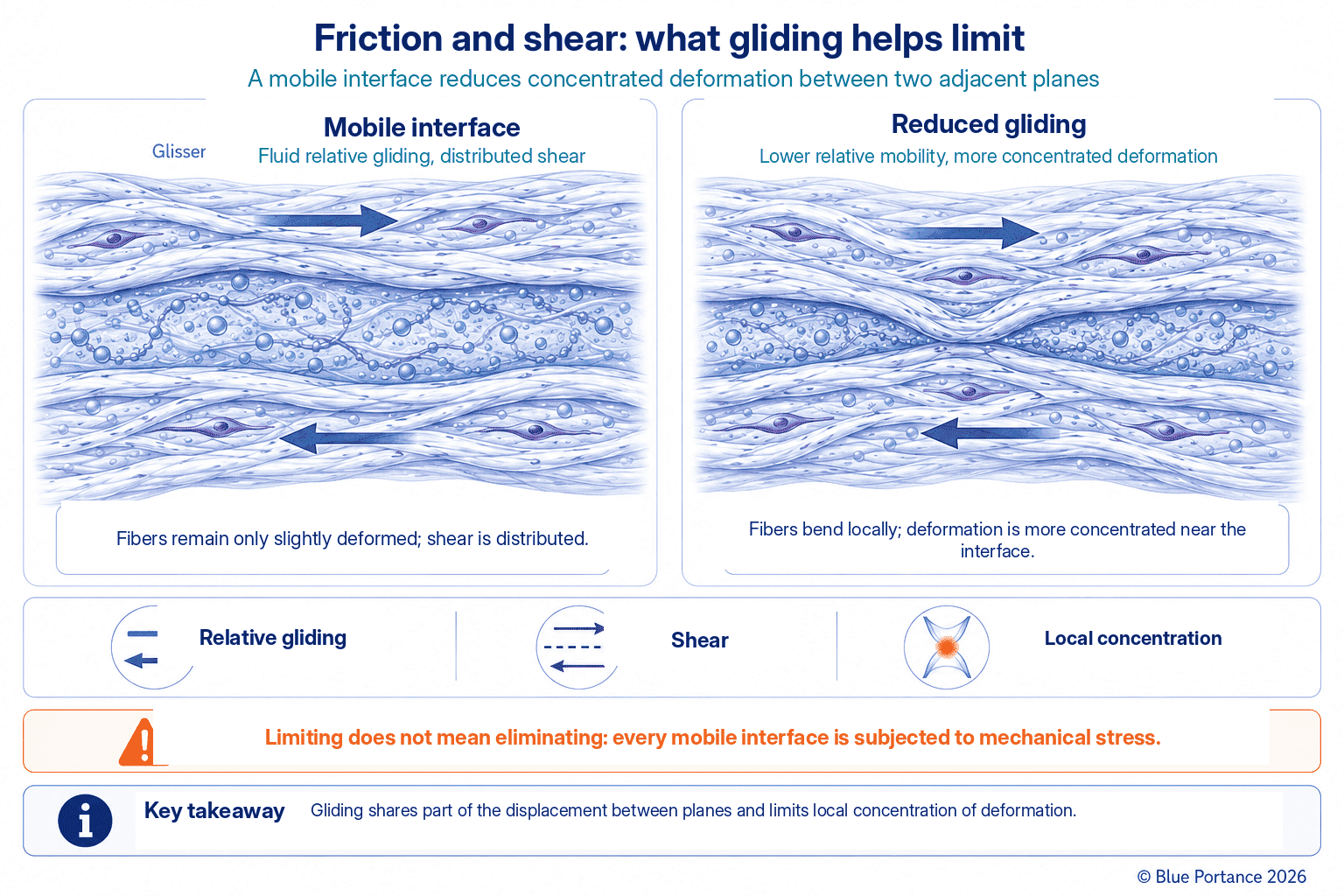
Interfaces distribute displacements across several planes instead of concentrating them at a single boundary. Water and hyaluronan contribute, in particular, to the mobility of certain layers. Hyaluronan content, quantified in various human fasciae, varies according to location and the gliding function specific to each anatomical site (Fede et al., 2018). When this mobility decreases, gliding resistance and shear strain can thus increase locally. These notions must be distinguished from each other, as well as from the internal deformation of the tissue itself.
Distributing Movement Across Several Interfaces
When several mobile interfaces actually take part in the displacement, each can absorb part of the total deformation: an overall movement then translates into a small amount of gliding at each interface. If one interface loses its mobility, the distribution of movement among the other levels may be altered and become more concentrated. This can then shift stress onto an area that is not designed for it. This logic of distribution across several successive interfaces assumes that they actually take part in the movement. This is one of the reasons why the mobility of several superimposed layers — skin, superficial fascia, aponeurosis, muscle — may contribute more to tissue protection than the flexibility of a single isolated layer.
The finger flexor tendons illustrate this principle particularly clearly by combining two distinct systems. The synovial sheath provides gliding with a very low coefficient of friction, comparable to that of articular cartilage. This gliding relies on lubricants bound to the tendon surface — hyaluronan, phospholipids, and a lubricating proteoglycan, lubricin (Sun et al., 2013). The fibrous pulleys, for their part, hold the tendon against the bone and prevent it from “bowstringing” during flexion. Some of them, notably the A2 and A4 pulleys, play a more decisive biomechanical role than the others in this function.
Two Combined Systems, a Fragile Balance
Within a very small space, this arrangement thus combines the three functions described in this article: maintaining anatomical position, low-resistance gliding, and distributing stress across several pulleys rather than onto a single point. A damaged synovial sheath or a ruptured or insufficient pulley, however, disrupts this balance. The tendon may then move abnormally during flexion, which redistributes the load less favorably along the rest of the tendon’s course.
5. Protection Is a Dynamic Property
A tissue is not protected because it does not move, but because movements and loads remain compatible with its capacities. Protection thus depends on variability, recovery, and coordination between structures.
This idea runs counter to an intuitive but misleading notion that immobility is protective by default and in all circumstances. Over the long term, tissue protection generally does not rest on permanent immobility, but on movements and loads compatible with the tissue’s capacities. A tissue held in the same position for a long time loses the micro-variations in load and gliding that normally allow it to distribute stress across several areas over time. Temporary immobilization may nevertheless be necessary in certain situations — for example, during certain phases of healing — but it then follows a different logic, distinct from the dynamic protection described here.
Changing Support to Renew the Distribution of Stress
Changes in position or support can renew the distribution of loading when they actually modify the load applied to the area in question. Their effect thus depends on their direction, amplitude, and frequency, and on the available support surfaces. A small amplitude does not, therefore, by itself guarantee that an area is effectively offloaded. This notion of micro-movement is developed in detail in the next chapter.
In practical terms, this dynamic reading of tissue protection has a direct implication for sitting: the issue is not simply choosing the “softest possible” seat. Softness does not guarantee offloading, and firmness does not automatically mean concentrated pressure. In reality, geometry, sinking depth, stability, support surface, and mobility all interact to determine whether tissues can continue to make their internal adjustments — gliding, redistribution, micro-variations — rather than being locked into a single, prolonged configuration. This assumes, however, that these adjustments remain possible without imposing permanent instability. This reading of fascia as a dynamic protective organ is also the one synthesized by Lesondak (Lesondak, 2019).
Key Takeaways
- Fascia maintains anatomical relationships without having to make them rigid.
- Gliding distributes displacements across several interfaces.
- The connective environment accompanies nerves and vessels.
- Tissue protection is dynamic and collective.
Frequently Asked Questions
Does fascia protect a nerve?
Is an adhesion always visible?
Does stability require stiffness?
Does movement always protect tissues?
Scientific References
Cited References (1/2)
- Adstrum S, Hedley G, Schleip R, Stecco C, Yucesoy CA. Defining the fascial system. J Bodyw Mov Ther. 2017;21(1):173–177.
- Fede C, Angelini A, Stern R, Macchi V, Porzionato A, Ruggieri P, De Caro R, Stecco C. Quantification of hyaluronan in human fasciae: variations with function and anatomical site. J Anat. 2018;233(4):552–556.
- Guimberteau JC, Delage JP, McGrouther DA, Wong JKF. The microvacuolar system: how connective tissue sliding works. J Hand Surg Eur Vol. 2010;35(8):614–622.
- Nagita H, Wang C, Saigusa H, Hoshina K, Suhara M, Oshima M. Deformed popliteal artery due to highly flexed knee position can cause kinks, creating an unfavorable hemodynamic state. Circ J. 2024;88(3):351–358.
- Stecco C. Functional Atlas of the Human Fascial System. Elsevier; 2015.
Cited References (2/2)
- Sun YL, Zhao C, Jay GD, Schmid TM, An KN, Amadio PC. Effects of stress deprivation on lubricin synthesis and gliding of flexor tendons in a canine model in vivo. J Bone Joint Surg Am. 2013;95(3):e13.
- Topp KS, Boyd BS. Structure and biomechanics of peripheral nerves: nerve responses to physical stresses and implications for physical therapist practice. Phys Ther. 2006;86(1):92–109.
- Wang Y, Zhao C, Passe SM, Filius A, Thoreson AR, An KN, Amadio PC. Transverse ultrasound assessment of median nerve deformation and displacement in the human carpal tunnel during wrist movements. Ultrasound Med Biol. 2014;40(1):53–61.
- Lesondak D. Le Fascia. [Fascia.] Éditions Ressources Primordiales; 2019.
← Previous chapter: How Does Fascia Transmit Forces and Distribute Stress?
Back to the Fascia guide
Next chapter: Why Does Fascia Need Micro-Movements? →
Note: this content explains general mechanisms. It does not constitute a medical diagnosis or a therapeutic recommendation.
© Gil Ayache. The original concepts, biomechanical models, diagrams, terminology, graphic representations, foundational figures, original texts, and principles presented on this page are works protected by copyright. They are made available to Blue Portance under an intellectual property license agreement, without transfer of economic rights or of authorship.
