


This article is part of the Expert Guide “Fascia.”
Chapter 3 — Blue Portance Knowledge BaseHow Does Fascia Transmit Forces and Distribute Stress?
Summary — Fascia connects structures that generate, receive, and transmit forces. Its fibrous architecture can spread a load, change its direction, and share it among several tissues. This redistribution depends on attachments, fiber orientation, the mobility of interfaces, and muscle involvement. It must be distinguished from energy dissipation and from contact pressure.
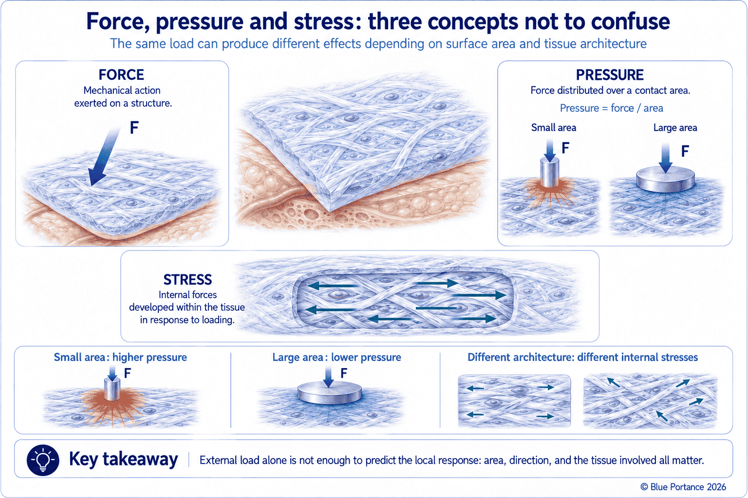
1. Force, pressure, and stress: three different concepts
A force describes a mechanical action. Pressure, for its part, relates that force to a contact surface. Stress, in turn, describes the internal forces per unit area within the tissue. A sitting pressure measured at the interface therefore does not directly reveal all internal stresses.
These three quantities differ in what they measure and where they measure it. Force is a global quantity, expressed in newtons, that says nothing about how it is distributed. Pressure, measured for example by a sensor mat placed under a seated user, corresponds to an average value over the area of each sensing cell, at the interface between the body and the seat. It therefore provides information only about that contact surface. Stress, by contrast, is defined within the tissue itself. It can thus vary considerably from one point to another, even beneath an area where the contact pressure measured at the surface appears uniform.
What imaging-based modeling shows
Models combining MRI and finite element analysis show that peak stresses and strains can occur in deep tissues, particularly muscle, without being directly inferable from the pressure measured at the interface alone (Linder-Ganz et al., 2007). Two devices producing different surface pressures can even maintain similar deep stress profiles (Boyle et al., 2020). Thus, two seats can show a fairly similar average pressure or surface pressure map while producing different deformations at depth. The shape of the ischial tuberosity, the thickness and properties of the soft tissues, and their capacity to glide all alter the compression and shear of muscle and fat. Interface pressure alone therefore cannot predict it.
The role of soft-tissue thickness
When sitting, the soft tissues are loaded between the support surface and bony prominences, particularly the ischial tuberosities. The geometry of these prominences, the thickness of the different layers, their mechanical properties, and their capacity to glide determine the distribution of internal stresses. Soft-tissue thickness alters this distribution, but its effect is not uniformly protective. Models show a complex relationship between fat thickness, body geometry, muscle atrophy, and internal stresses, with some configurations even showing an increase in deep stresses with body mass index (Elsner & Gefen, 2008) (Chen et al., 2022). The protective effect of a thicker fat layer therefore also depends on the shape of the bony prominences, the properties of muscle and fat, and how they deform under load.
2. Transmitting without transmitting everything
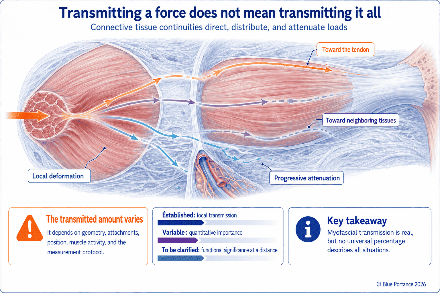
Muscle fibers insert into aponeuroses and septa; part of their force can be transmitted laterally through connective tissues (Huijing, 2003). Connections between synergistic muscles also allow local transfers.
Huijing proposes a “multilevel” reading of this transmission. The force generated by a muscle does not follow a single pathway. It is distributed simultaneously along the tendon, through myofascial connections with neighboring muscles, and via the connective tissue sheaths that link the muscle to adjacent compartments (Huijing, 2003). When a muscle develops force, most of it is transmitted to its tendon. A fraction may also be transmitted to the intermuscular septa and neighboring connective tissues, depending on the anatomical connections, joint position, and the activation state of the muscles involved. Subsequent studies have confirmed that closely neighboring synergistic muscles can exchange part of their force through these connective tissue linkages. This exchange thus takes place outside each muscle’s own tendon pathway (Maas & Sandercock, 2010).
A transmission that attenuates and is shared
This transmission attenuates and is shared. It therefore depends on joint position, muscle activity, and anatomical boundaries. A systematic review emphasizes that intermuscular transmission chains do indeed exist. Their overall mechanical contribution nevertheless remains difficult to quantify unambiguously, as it depends so heavily on experimental conditions — joint angle, contraction level, muscles involved (Krause et al., 2016). It would therefore be an overstatement to present this transmission as systematically dominant over the classic tendon pathway. It is a real and documented mechanism, but an additional one, which does not replace known tendon anatomy — an interpretive caution shared by Lesondak’s synthesis (Lesondak, 2019).
3. Distributing a load in space
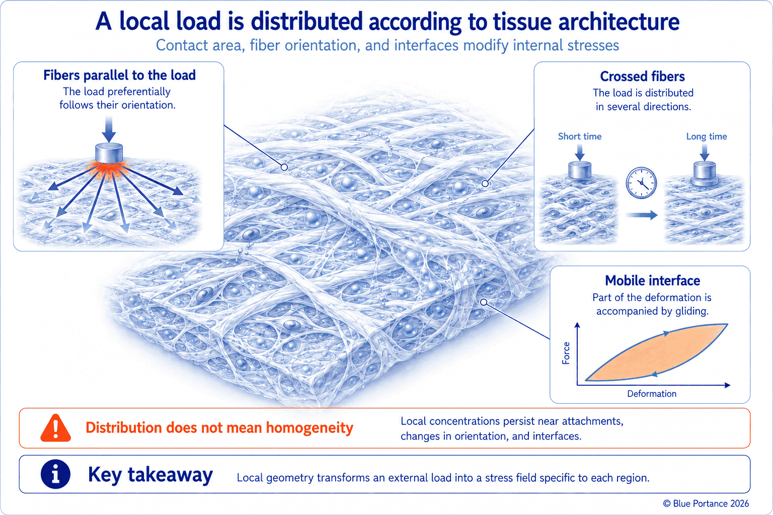
A large support surface generally reduces average pressure, but the internal distribution also depends on the thickness, stiffness, and shape of the tissues. The assembly formed by skin, adipose tissue, fascia, and muscle contributes in particular to this redistribution. The respective contribution of each layer thus depends on its geometry, its mechanical properties, and its interfaces, with the fascial layers in particular transmitting tension in several directions. Increasing the contact area can reduce average pressure. But if the load remains concentrated around a bony prominence, if the tissues glide poorly, or if substantial shear develops, deep stresses can remain high: the spreading visible at the surface therefore does not guarantee an equivalent redistribution throughout the full thickness of the tissues.
A useful image: the stretched membrane
In a simplified membrane model, a tensioned fascial sheet can be compared, with caution, to an elastic membrane on which a weight rests. The load is thus not borne solely at the point of contact, but is distributed along the tensioned fibers to their peripheral anchoring points. The more tensioned the membrane, and the more its fibers are oriented so as to converge toward the loaded area, the more effective this spreading. Conversely, a slack or loosely tensioned tissue concentrates the load more at the point of application, with less redistribution toward the periphery. This image nevertheless remains a simplification. Unlike an ideal isotropic membrane, real fascia has a preferential fiber orientation, so that the redistribution is itself anisotropic and favors certain axes.
Anisotropy and the biotensegrity model
Anisotropy guides the load along certain orientations. A change in gliding or in fiber organization — after a scar, for example — may alter these pathways and the local distribution of stresses.
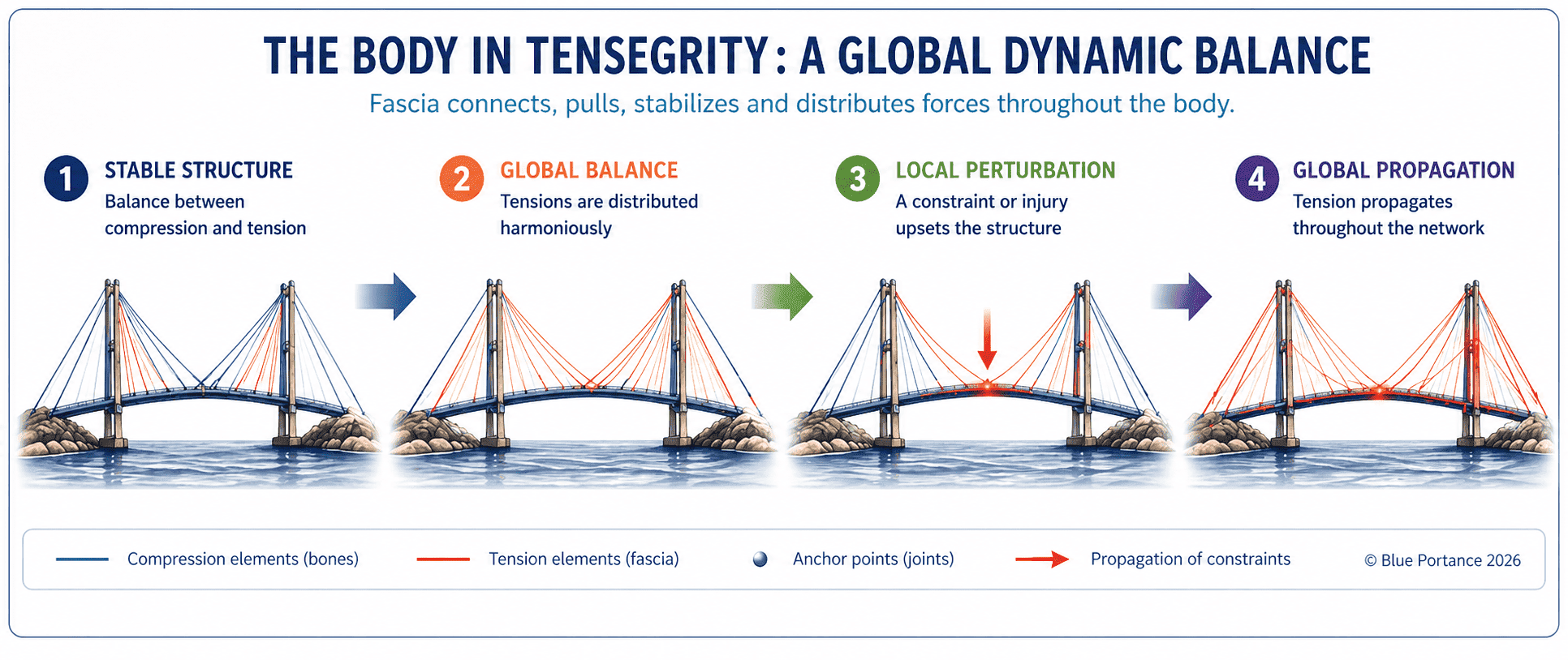
Biotensegrity proposes reading the body’s mechanical balance as a system of tension and compression distributed across a continuous network of connective tissues, rather than as a stack of rigid segments resting on one another. This framework thus helps visualize how a load can be redistributed within a connected structure. It nevertheless remains an interpretive model: useful for thinking about certain stress-distribution phenomena, it does not constitute a validated experimental demonstration of all its implications (Bordoni & Myers, 2020).
4. Redistributing over time
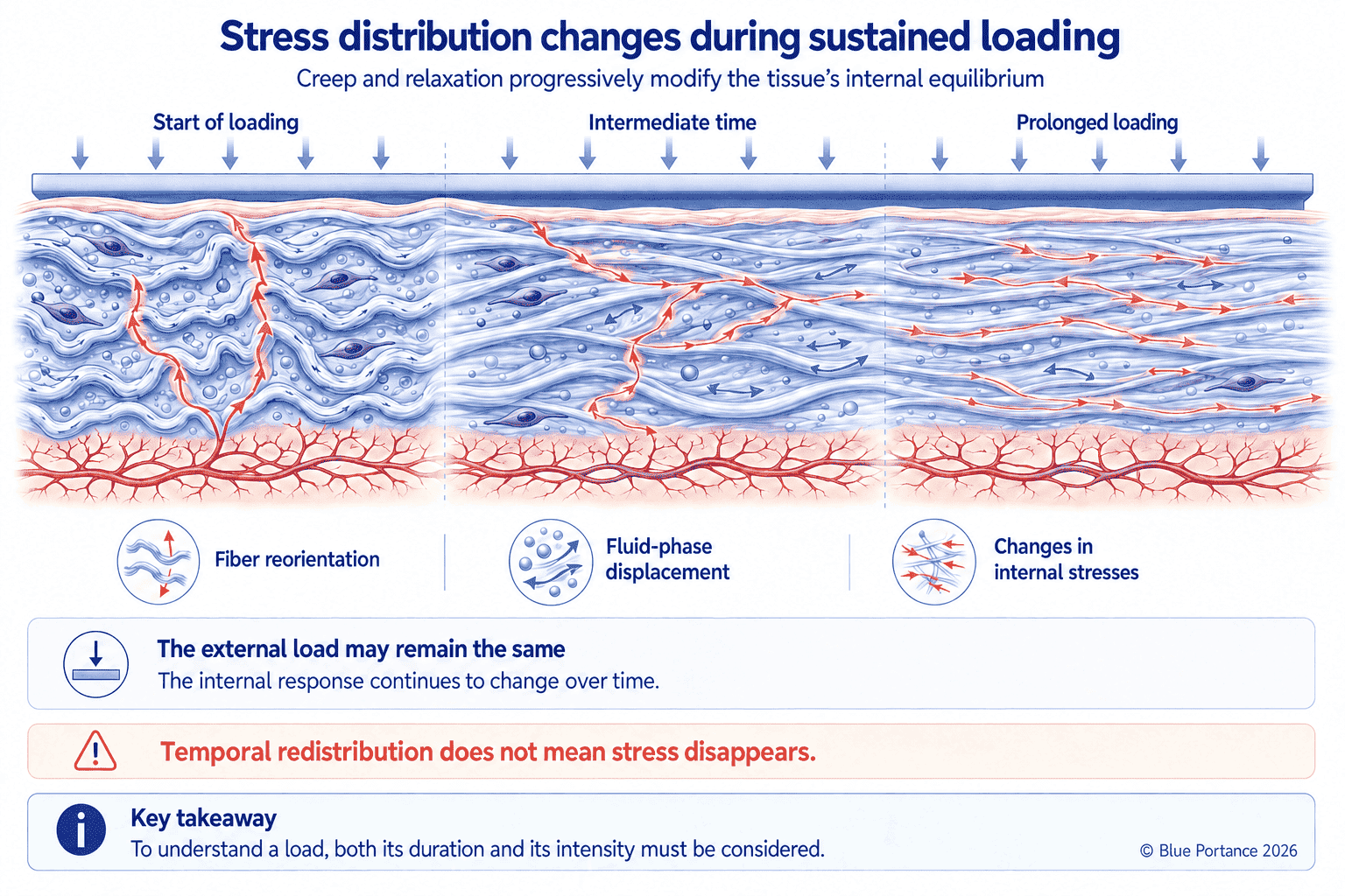
Viscoelasticity gradually alters the distribution: creep, stress relaxation, and fluid movement change the share of the load carried by each component. The same external pressure therefore does not imply a constant internal state throughout the exposure.
This temporal redistribution, described in Chapter 2 at the scale of an isolated tissue, becomes more complex when several superimposed tissues are involved. Beneath the ischial tuberosities, the load simultaneously engages skin, fat, fascia, and muscle. Some layers are compressed, others shift laterally, while their interfaces can glide relative to one another. In vivo MRI measurements confirm this complexity, including measurable sliding of the gluteus maximus relative to the ischial tuberosity under load (Zappalá et al., 2024).
Under sustained load, the viscoelastic deformation of fat and muscle evolves over time. The distribution of stresses among the different layers can therefore change, depending on their mechanical properties, their geometry, and their capacity to glide. Redistribution thus depends less on any single layer than on the mechanical organization of the whole. These mechanical changes may contribute to a gradual change in the sensation of support after several minutes of immobility, without being sufficient on their own to explain the discomfort felt, which also depends on perfusion, innervation, local temperature, and individual sensitivity.
5. Spreading and dissipation are not the same thing
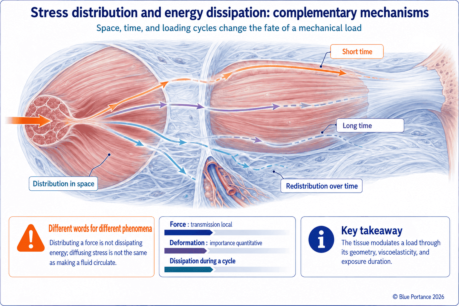
Spreading, or distribution, describes a spatial redistribution. Dissipation describes a transformation of energy during deformation. A tissue can thus distribute a force without dissipating much energy, or dissipate energy in an area that remains heavily loaded.
These two concepts answer different questions. Spreading answers the question “Where does the load go?”: it describes how a force is distributed among several areas or several tissues. Dissipation, for its part, answers the question “What happens to the energy?”: it describes the portion of mechanical energy that is not returned when the tissue goes back to its initial state, mainly converted into heat, as discussed in Chapter 2 in connection with hysteresis.
A highly elastic tissue can thus effectively spread a load over a large area — good spreading — while returning almost all of the stored energy during unloading — low dissipation. Conversely, a highly damped tissue can dissipate a great deal of energy without effectively distributing the load in space, if its contact area remains localized. Confusing these two properties leads to erroneous interpretations: a “sinking-in” sensation or perceived cushioning in a seat does not, in itself, guarantee better spatial distribution of stresses. A very soft foam can thus absorb part of the energy and give a sensation of cushioning, while letting the bony prominences sink in and pressures concentrate locally. Conversely, a low-dissipation structure can effectively spread a load over a larger area. Damping, distributing, and allowing weight-bearing to alternate are therefore three distinct mechanical functions.
Key Takeaways
- External pressure and internal stress are not equivalent.
- Fascia contributes to the local and regional transmission of forces.
- Distribution depends on architecture, attachments, and gliding.
- Spatial redistribution and energy dissipation must be distinguished.
Frequently Asked Questions
Does low pressure guarantee the absence of stress?
Can fascial tension act at a distance?
Does fascia absorb shocks?
Scientific References
Cited References (1/2)
- Boyle CJ, Carpanen D, Pandelani T, Higgins CA, Masen MA, Masouros SD. Lateral pressure equalisation as a principle for designing support surfaces to prevent deep tissue pressure ulcers. PLOS One. 2020;15(1):e0227064.
- Chen Y, Shen Y, Wang K, et al. Mechanical analysis of deep tissue injury during sitting in patients with spinal cord injury via parametric finite element model. Biomech Model Mechanobiol. 2022;21(5):1573–1584.
- Elsner JJ, Gefen A. Is obesity a risk factor for deep tissue injury in patients with spinal cord injury? J Biomech. 2008;41(16):3322–3331.
- Fung YC. Biomechanics: Mechanical Properties of Living Tissues. Springer; 1993.
- Linder-Ganz E, Yarnitzky G, Yizhar Z, Siev-Ner I, Gefen A. Assessment of mechanical conditions in sub-dermal tissues during sitting: a combined experimental-MRI and finite element approach. J Biomech. 2007;40(7):1443–1454.
- Yahia L, Pigeon P, DesRosiers EA. Viscoelastic properties of the human lumbodorsal fascia. J Biomed Eng. 1993;15(5):425–429.
Cited References (2/2)
- Zappalá S, Keenan BE, Marshall D, Wu J, Evans SL, Al-Dirini RMA. In vivo strain measurements in the human buttock during sitting using MR-based digital volume correlation. J Biomech. 2024;163:111913.
- Huijing PA. Muscular force transmission necessitates a multilevel integrative approach. Exerc Sport Sci Rev. 2003;31(4):167–175.
- Maas H, Sandercock TG. Force transmission between synergistic skeletal muscles through connective tissue linkages. J Biomed Biotechnol. 2010;2010:575672.
- Krause F, Wilke J, Vogt L, Banzer W. Intermuscular force transmission along myofascial chains: a systematic review. J Anat. 2016;228(6):910–918.
- Bordoni B, Myers T. A Review of the Theoretical Fascial Models: Biotensegrity, Fascintegrity, and Myofascial Chains. Cureus. 2020;12(2):e7092.
- Lesondak D. Le Fascia. [Fascia.] Éditions Ressources Primordiales; 2019.
← Previous chapter: How Does Fascia Behave Under Stress?
Back to the Fascia guide
Next chapter: What Role Does Fascia Play in Tissue Dynamics and Protection? →
Note: this content explains general mechanisms. It does not constitute a medical diagnosis or a therapeutic recommendation.
© Gil Ayache. The original concepts, biomechanical models, diagrams, terminology, graphic representations, foundational figures, original texts, and principles presented on this page are works protected by copyright. They are made available to Blue Portance under an intellectual property license agreement, without transfer of economic rights or of authorship.
