


6. Coccyx Morphology and Mobility: A Multifactorial Mechanical Vulnerability
Knowledge Base – Coccydynia Expert Guide
Beyond functional and neurological mechanisms, each individual’s own anatomy defines a background mechanical vulnerability that can now be classified.
The morphology of the coccyx is neither uniform nor neutral in the general population. Anatomical and radiological data show substantial interindividual variability, which determines how the mechanical stress of sitting, movement, and trauma is absorbed or, on the contrary, concentrated.
This morphological diversity defines a background mechanical vulnerability, neither pathological in itself nor systematically painful, but one that influences how the system responds to environmental stress.
1. The reference morphological classification
The founding work of Postacchini and Massobrio (1983) proposed a morphological classification of the coccyx into four main types, based on the curvature and orientation of the coccygeal segments (Postacchini & Massobrio, 1983).
- Type I: coccyx slightly curved forward, extending the sacrum harmoniously.
- Next, Type II: more pronounced ventral curvature, tip pointing into the pelvis.
- Then, Type III: sharp anterior angulation between two segments.
- Finally, Type IV: subluxation or intersegmental instability.
Identifying these types relies notably on dynamic sitting/standing radiographs, as standardized by Maigne and Tamalet (1996) (Maigne & Tamalet, 1996).
This approach showed a correlation between certain morphologies (II, III, IV) and a higher incidence of coccydynia, including in the absence of any lesion visible on static imaging (Maigne & Tamalet, 1996; Nathan et al., 2010).
2. Anatomical predisposition and response to trauma
For the same trauma — a comparable fall, a similar childbirth, identical sitting stress — some individuals develop persistent chronic pain, while others recover spontaneously.
Pain then appears as the meeting point between:
- a specific bony architecture (morphological background), and
- an environmental mechanical stress (sitting, repetition, impact),
- which the system can no longer absorb (adaptive dysfunction).
3. Toward a broader morphological reading
Clinical data have broadened the morphological reading to six profiles, including retroverted or lateralized forms, and configurations associated with spicules (Nathan et al., 2010; Garg & Ahuja, 2021).
These morphotypes are not isolated causes. They must be read as mechanical vulnerability factors, whose expression depends on dynamic behavior.
- A morphotype is a constitutional anatomical configuration: interindividual variation in curvature, angulation, or alignment. It defines a background, not a diagnosis.
- A lesion, by contrast, is an acquired, objectifiable alteration: fracture, dislocation, unstable subluxation, post-traumatic instability.
- Without this distinction, a mistaken conclusion becomes possible: “I have a Type III → I am ill.” In reality, the correct doctrine is: morphology defines a background; a lesion corresponds to a breakdown of the adaptive balance.
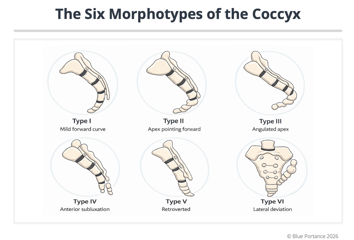
Morphological variations (Types I to VI). Morphology defines a mechanical background of vulnerability; clinical expression depends on dynamic mobility and on the stress of sitting (Postacchini & Massobrio, 1983; Maigne & Tamalet, 1996).
Summary table of the 6 morphotypes
| Type | Anatomical description | Mechanical characteristics | Potential mechanical relevance |
|---|---|---|---|
| Type I | Gentle curvature, aligned with the sacrum | Good stress dissipation | Usually compatible with good adaptation |
| Type II | Pronounced ventral curvature | Stress concentration when sitting | Relevant if mobility is reduced |
| Type III | Anterior segmental angulation | Localized point of mechanical conflict | Relevant if the angulation becomes a point of conflict |
| Type IV | Sacrococcygeal or intercoccygeal subluxation | Dynamic instability (sitting/standing) | Instability to look for on dynamic radiographs |
| Type V | Retroversion ± bony spicule | Chronic tissue irritation | Relevant if a spicule irritates the soft tissues |
| Type VI | Lateral deviation / asymmetric alignment | Asymmetric stress | May explain atypical or referred pain |
These profiles are neither a diagnosis nor a fate. They represent mechanical vulnerability factors, whose clinical expression depends on actual mobility, the condition of the soft tissues, and repeated stress.
4. Segmental biomechanics: from morphotype to mechanical behavior
Speaking of the overall “shape” of the coccyx is useful for classification, but insufficient for understanding its mechanics.
The coccyx is a terminal hinge made up of 3 to 5 segments, linked by intercoccygeal joints, and articulated with the sacrum (sacrococcygeal joint). Its mechanics are therefore not those of a single block.
Each morphotype influences:
- the location of the center of rotation during movement,
- the existence of a segmental angulation point,
- the presence of a stress concentration zone when sitting,
- and the way forces are dissipated… or focused.
For example, a Type III is not simply “more curved.” It is a segmental angulation that can become a point of mechanical conflict. A Type IV adds a dimension of dynamic instability, generally better demonstrated with functional (sitting/standing) imaging (Maigne & Tamalet, 1996).
- We no longer read only: “Here are the 6 shapes.”
- We read: “Here is how each architecture can behave dynamically.”
5. Physiological mobility: hypermobility vs hypomobility
In reality, the coccyx is not only a shape: it is a dynamic segment. Dynamic sitting/standing radiographs have shown a physiological mobility, generally described as a flexion of about 5 to 25 degrees, hypermobility being defined beyond 25 degrees, underscoring its role as a terminal shock absorber (Maigne & Tamalet, 1996).
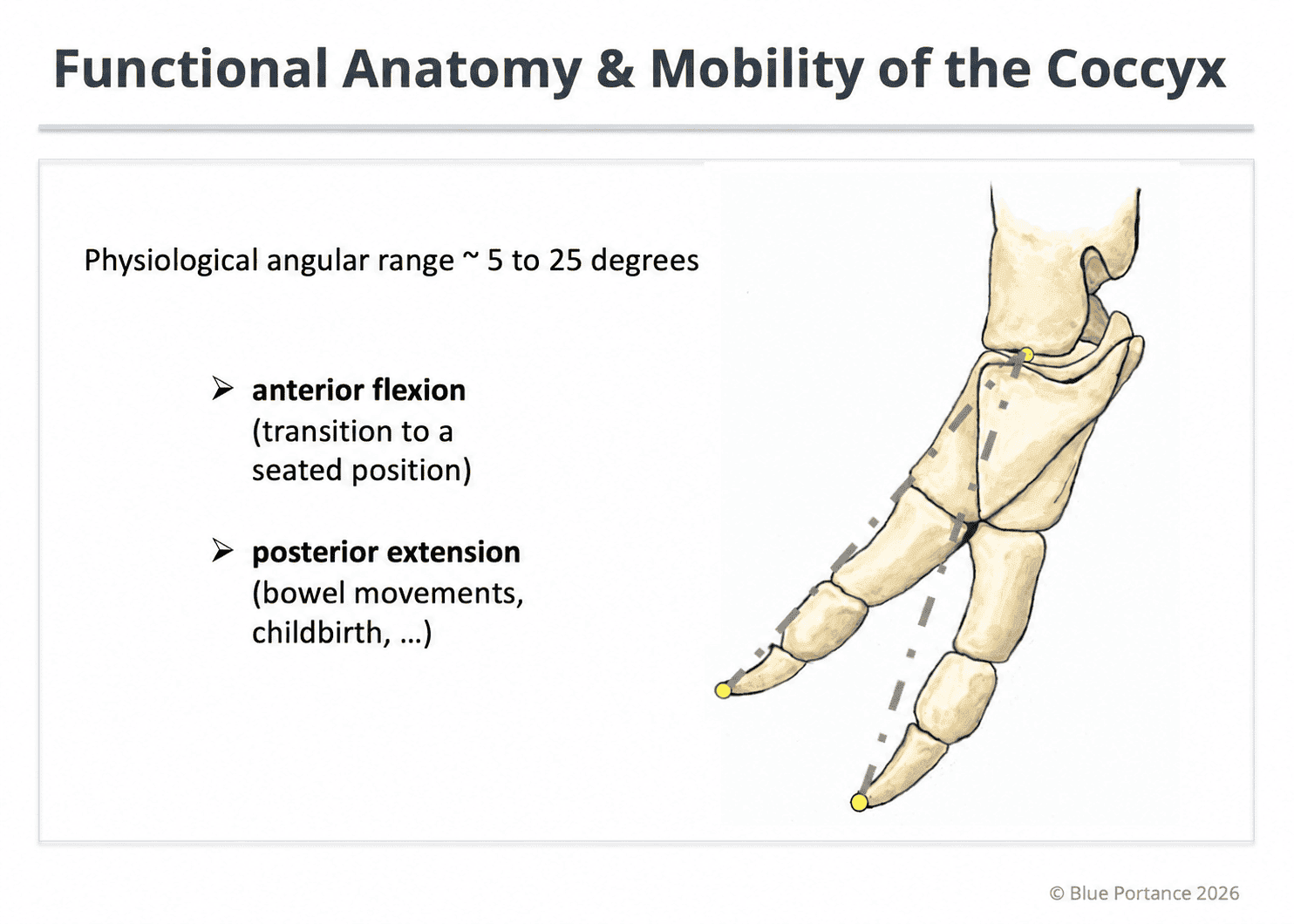
Illustration of the normal angular range in flexion-extension (about 5–25°) observed on dynamic sitting/standing imaging. The coccyx acts as an adaptive terminal hinge of the spine. Hypermobility (excessive angulation) and hypomobility (stiffening) can both impair stress dissipation and promote abnormal transmission of loads to the soft tissues (Maigne & Tamalet, 1996).
Two opposite drifts can produce pain:
- Hypermobility: excessive angulation when sitting, segmental instability, repeated micro-trauma, local nociceptive activation — frequently described in presentations associated with Types III–IV when they are unstable (Maigne & Tamalet, 1996; Nathan et al., 2010).
- Hypomobility: adaptive locking, loss of terminal shock absorption, more direct transmission of loads. Often less visible on static imaging, but clinically relevant (post-traumatic / protective stiffening).
The doctrinal consequence: the same morphology can be asymptomatic if it remains mobile, painful if it stiffens, and painful if it becomes unstable. Pain depends on the interaction: Morphology × Mobility × Environmental stress.
Simple examples:
- Type II mobile → shock absorption preserved; Type II rigid → load concentration.
- Type III stable → adaptation possible; Type III unstable → repeated micro-trauma.
- Types V–VI: retroversion / asymmetry → mechanical vectors that can increase tissue conflict or referred pain depending on the context.
6. Interaction with the soft tissues: the clinical “so what”
The coccyx is not an isolated bone. It sits at a crossroads of attachments and tension: pelvic floor, sacrotuberous ligament, gluteus maximus, posterior fascial convergence. Consequently, any variation in morphology or mobility changes tissue tension.
In practice:
- If hypermobile: repeated ligamentous traction, irritation of the muscle attachments, local micro-inflammation, peripheral sensitization.
- If hypomobile: loss of shock absorption, compression of the posterior tissues, loss of fascial gliding, defensive hypertonia of the pelvic floor.
In both cases, the pain is not “purely bony”: it becomes myofascial and neuromechanical. Morphology does not directly cause pain; it changes how forces are transmitted to the soft tissues, which can trigger local inflammation, reflex contraction, and a vicious cycle of pain (Nathan et al., 2010; Garg & Ahuja, 2021).
Doctrinal conclusion
- Coccygeal morphology is neither a diagnosis nor a direct cause: it defines a mechanical background.
- Pain emerges from the interaction between bony architecture (morphotype), dynamic behavior (hyper/hypomobility), environmental stress (sitting, trauma, repetition), and the response of the soft tissues (muscles, ligaments, fascia).
- Coccydynia is not only a bone problem: it is a dynamic interaction between a terminal hinge and the tissues around it.
- Ultimately, the clinical goal is to identify the dominant mechanism (instability, stiffening, tissue conflict) to guide assessment and strategy.
- Postacchini F, Massobrio M. Idiopathic coccygodynia. J Bone Joint Surg Am. 1983.
- Maigne JY, Tamalet B. Standardized radiologic protocol for the study of common coccygodynia and characteristics of the lesions observed in the sitting position. Spine. 1996;21(22):2588–2593.
- Maigne JY, Lagauche D, Doursounian L. Instability of the coccyx in coccydynia. J Bone Joint Surg Br. 2000;82(7):1038–1041.
- Nathan ST, Fisher BE, Roberts CS. Coccydynia: a review of pathoanatomy, aetiology, treatment and outcome. J Bone Joint Surg Br. 2010;92(12):1622–1627.
- Garg B, Ahuja K. Coccydynia — A comprehensive review on etiology, radiological features and management options. J Clin Orthop Trauma. 2021;12(1):123–129.
© Gil Ayache. The concepts, diagrams, terminology, and principles presented on this page constitute a work protected by copyright. They are made available to Blue Portance under an intellectual property license agreement, without transfer of economic rights or of authorship.
