


1. The body as an open system
In biology, an open system is an organization that continuously exchanges with its environment. This exchange involves both matter and energy. By contrast, a closed system would exchange only energy, and an isolated system would exchange nothing at all. The living body falls into the first category.
As early as 1968, the biologist Ludwig von Bertalanffy formalized this idea in his general systems theory. A living organism cannot be understood as a static object. Instead, it maintains itself precisely by remaining open to exchanges with its surroundings (Bertalanffy, 1968). In fact, this openness is not one option among others: it is the very condition of its existence.
This guide takes a broad view of these exchanges: matter and energy, but also information. Here, the latter term is understood in the biological sense. It refers in particular to any signal capable of altering the state or activity of a living system (Hoke et al., 2021). However, sensory and nerve flows as such will be covered in a separate sub-guide.
Describing the body solely through its anatomy — its bones, its muscles, its joints — is not enough. Indeed, it is a bit like photographing a river without showing the flowing water. The shape remains recognizable, while part of the components that sustain it is continuously transferred, transformed or renewed.
A living organization far from equilibrium
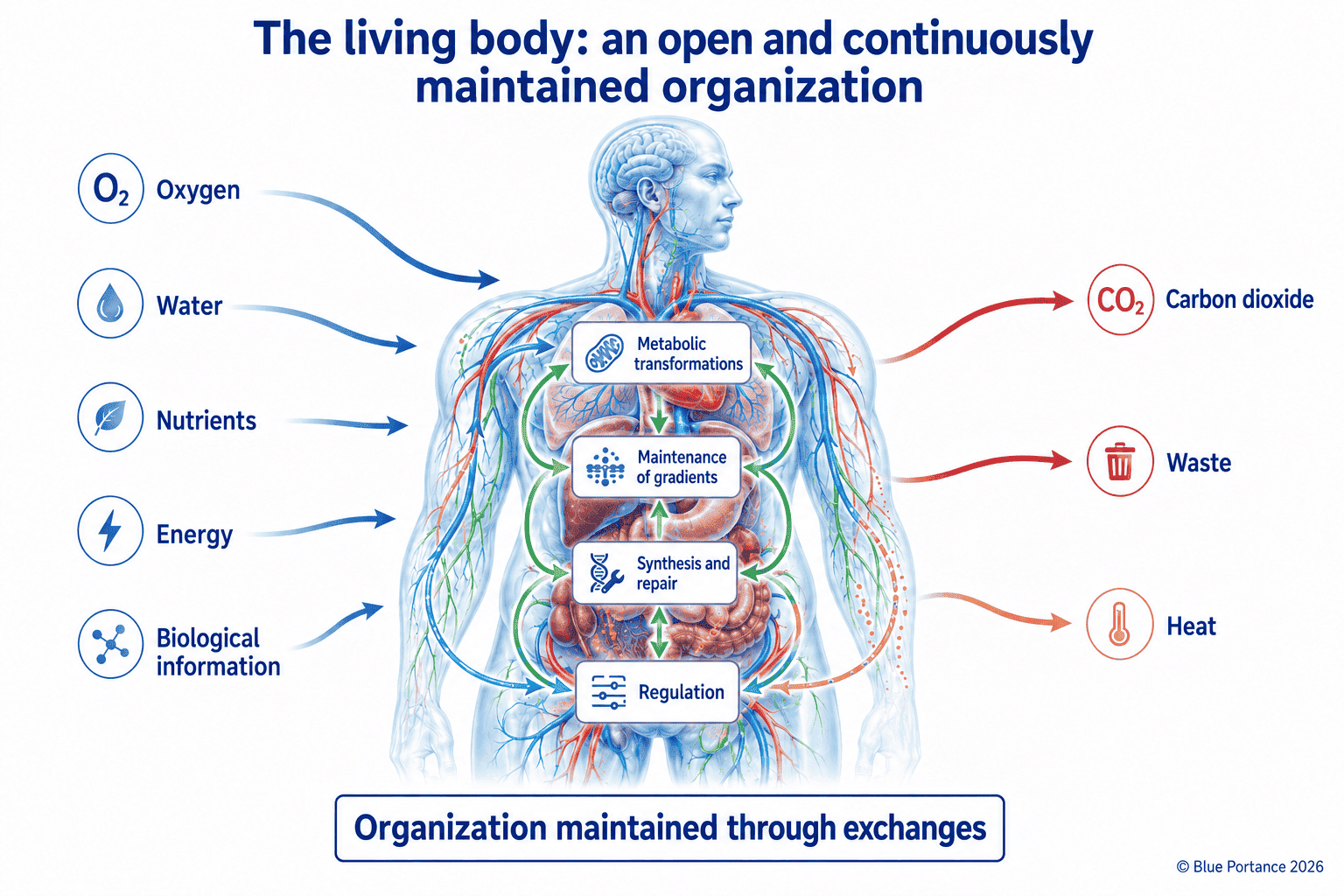
In 1944, in What Is Life?, the physicist Erwin Schrödinger put forward an idea that has since become classic. A living organism, he argued, maintains its organization through continuous exchanges with its environment. These exchanges involve both matter and energy (Schrödinger, 1944). An isolated system, by contrast, spontaneously evolves toward a state of thermodynamic equilibrium in which usable gradients fade away.
2. Inputs, transformations and outputs: the exchanges that sustain metabolism
In practice, this exchange takes the form of three simultaneous movements. First, the body takes in matter and energy — oxygen, water, nutrients. Next, it transforms them through successive metabolic reactions. It then eliminates the products of this transformation — carbon dioxide, heat, nitrogenous waste.
These transformations rely in particular on adenosine triphosphate, or ATP. This molecule is the main immediately usable form of chemical energy for many cellular functions. Among other things, it powers the maintenance of ion gradients across membranes. It is also involved in active transport and muscle contraction. It likewise supports signaling, as well as the synthesis and repair of tissue components (Nelson & Cox, 2021).
Metabolism thus refers to the organized set of reactions that transform matter and energy within the organism. This transformation operates from the molecular scale up to the scale of tissues. It classically has two complementary sides. Catabolism breaks down complex molecules to release their energy. Conversely, anabolism uses this energy to synthesize the components needed for tissue growth and renewal. In short, without this metabolic activity, the organism could not sustainably maintain its gradients, its cellular functions or its physiological organization.
This metabolism is not uniform, however: its intensity varies greatly from one organ to another. Expressed per kilogram of tissue, for example, it is far higher in the heart and kidneys than in resting skeletal muscle. Adipose tissue, by contrast, consumes comparatively very little energy (Wang et al., 2010). This metabolic heterogeneity at the organ level foreshadows the diversity of rhythms also found at the molecular and cellular levels. The next chapter will explore this diversity.
3. The continuous renewal of components
This flow is not limited to the supply of energy. It also involves the material renewal of the body’s components themselves. Proteins, membrane lipids and even some components of the extracellular matrix are continuously broken down and then resynthesized. These renewal rates nevertheless vary greatly from one molecule and one tissue to another.
Three nested levels of renewal
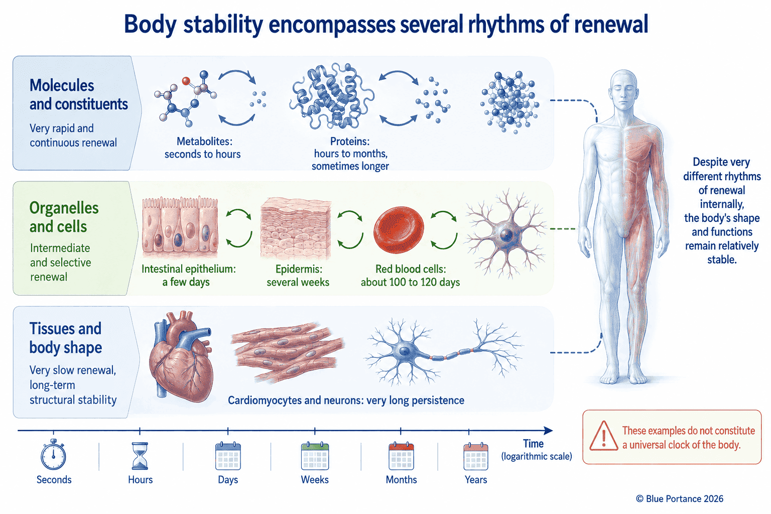
This renewal is also organized on several nested levels, which should be distinguished. For example, a molecule can be broken down and resynthesized without the organelle it belongs to ceasing to function. Likewise, an organelle can itself be renewed without the cell that houses it disappearing. A cell, for its part, may be replaced within a few days, depending on the tissue. Or, conversely, it may persist throughout an individual’s entire life. This, however, says nothing about the renewal rate of its own internal molecules. The macroscopic stability of a tissue — the stability visible to the naked eye — therefore tells us nothing about the persistence of its cells. Nor does it tell us anything about the persistence of their molecular components. These are three distinct scales, each evolving at its own pace.
These three scales can be illustrated with a few common examples. Circulating red blood cells are renewed in roughly a hundred days. They are then eliminated and replaced by new cells produced by the bone marrow. The epithelium lining the intestine renews itself in barely a few days. It is thus one of the most rapidly replaced tissues in the body. By contrast, the outer layer of the epidermis takes several weeks to renew itself completely (Sender & Milo, 2021). In other words, these very different cellular rhythms say nothing about the rate of molecular renewal. This holds for every tissue, whatever its cells.
Highly unequal renewal rates
These rhythms are spread across very different time scales. For example, energy molecules and certain metabolites are renewed within seconds to hours. Many cellular proteins, for their part, have variable lifespans, ranging from a few hours to several months depending on the tissue. Meanwhile, blood cells and epithelial cells are replaced within days to months. By contrast, some components of the extracellular matrix persist for several months, or even several years.
This heterogeneity calls for correcting a common misconception. Contrary to a fairly widespread image, the body does not renew all of its matter at regular intervals. Some structures are, on the contrary, remarkably stable. Certain proteins in the nucleus of the eye’s lens, called crystallins, are extremely stable. They show virtually no detectable turnover after their formation (Lynnerup et al., 2008). Similarly, one study focused in particular on the human occipital cortex. Some neurons there had an age comparable to that of the individual. The surrounding non-neuronal cells, on the other hand, had been renewed (Spalding et al., 2005). In the rat brain, structural proteins showed exceptionally long lifespans. These included, in particular, certain components of nuclear pore complexes and certain histones. They nevertheless retained slow but measurable exchange (Toyama et al., 2013).
This continuous yet heterogeneous renewal explains an apparent paradox. A bodily structure can remain visually stable for years. Yet a large share of its components has been transformed or replaced in the meantime. These rhythms vary widely from one molecule and one tissue to another. The shape persists, while part of its matter is continuously transferred, transformed or renewed.
Molecular renewal and cellular persistence: the example of muscle
Skeletal muscle clearly illustrates this distinction between scales. A large proportion of its fibers are post-mitotic cells. These cells persist for years, or even throughout the individual’s life, without cell division. Yet this cellular persistence does not mean molecular stability. In mice, quantitative proteomics studies have measured highly heterogeneous protein half-lives. These measurements concerned skeletal muscle tissue. For example, some proteins were renewed within a few hours, others over several months (Rolfs et al., 2021). This finding, obtained in animals, illustrates the general principle rather than providing a direct measurement in humans.
Heart muscle offers a comparable, though even more nuanced, example. Contrary to a long-held belief, human cardiomyocytes do renew themselves, albeit very slowly. Moreover, their annual renewal rate declines with age. Specifically, it falls from about 1% per year around age 25 to less than 0.5% around age 75. Fewer than half of an individual’s cardiomyocytes are thus replaced over an entire lifetime (Bergmann et al., 2009). A long-lived muscle cell can therefore renew a large part of its molecular content and even, very slowly, renew itself. However, this does not undermine the apparent stability of the tissue it forms.
4. Anatomical structure and living process: an essential distinction
This distinction between structure and process is central to this guide. An anatomical structure describes a shape at a given moment — a bone, a muscle, a joint. A living process, by contrast, describes what is continuously happening within and around that shape. More precisely, exchanges, transformations and renewal form its fabric.
According to one theoretical framework of biological autonomy, the organism behaves as an open, self-maintaining system. It produces and maintains its own components over time. This organism therefore does not function as a fixed assembly of parts (Moreno & Mossio, 2015). In other words, anatomy describes a geometry; physiology describes an activity. Both perspectives are necessary, but neither can replace the other.
5. Homeostasis: a dynamic balance, not immobility
It was the physiologist Walter Cannon who introduced the term homeostasis in 1929. He used it to describe the organism’s ability to maintain the relative constancy of its internal environment — temperature, pH, various concentrations. This constancy persists despite variations in its environment (Cannon, 1929).
This constancy, however, should not be confused with an absence of movement. On the contrary, it results from active and continuous regulation. This regulation constantly adjusts inputs and outputs to offset disturbances. Homeostasis is therefore a maintained balance, not a fixed state. It is thus distinct from the material renewal of tissues (Chapter 3) and from their adaptation to mechanical stresses, which follow different logics and time scales.
When a variable drifts away from its usual range, corrective mechanisms kick in to bring it back within that range. These mechanisms include, among others, vasodilation, shivering and hormonal secretions. This regulation can be observed, for example, in the maintenance of body temperature or blood glucose. Homeostasis therefore does not lock any value in place. Rather, it keeps variables within limits compatible with the organism’s functioning, through constant corrective activity.
6. What this changes for seated posture
Physiology firmly establishes one point: living systems maintain themselves through continuous exchanges, not through immobility. This general finding does not, however, establish that any particular seating device would by itself improve cellular function. The link between the two remains conceptual. It will be clarified chapter by chapter. As it progresses, this guide will address the extracellular matrix, mechanical stresses and then the seated position itself.
This first chapter thus mainly sets out a principle for interpretation. Specifically, maintaining a shape or a posture does not mean an absence of internal activity. A seat should therefore not be assessed solely on its ability to maintain a predefined alignment. The mechanical conditions it maintains over time must also be considered. Account must also be taken of the freedom left to the person to spontaneously change their posture and their support points.
The following chapters detail what these exchanges concretely involve: the internal environment of cells, then the extracellular matrix. They also describe the physical mechanisms — diffusion, osmosis, filtration — that enable the transfer of matter within tissues.
Ultimately, the living body appears stable because it maintains its organization, not because it remains still. This organization depends on constant exchanges of matter and energy. In addition, these exchanges are sustained at heterogeneous rhythms, depending on the molecules, cells and tissues involved. The following articles in this guide will each take up one of these mechanisms to clarify how it works. Ultimately, they will spell out its implications for the seated position.
Scientific References
Open system, metabolism and homeostasis
- Bertalanffy, L. von. (1968). General System Theory: Foundations, Development, Applications. New York: George Braziller.
- Cannon, W. B. (1929). Organization for physiological homeostasis. Physiological Reviews, 9(3), 399–431.
- Hoke, K. L., Zimmer, S. L., Roddy, A. B., Ondrechen, M. J., Williamson, C. E., & Buan, N. R. (2021). Reintegrating biology through the nexus of energy, information, and matter. Integrative and Comparative Biology, 61(6), 2082–2094.
- Moreno, A., & Mossio, M. (2015). Biological Autonomy: A Philosophical and Theoretical Enquiry. Dordrecht: Springer.
- Nelson, D. L., & Cox, M. M. (2021). Lehninger Principles of Biochemistry (8th ed.). New York: W. H. Freeman.
- Schrödinger, E. (1944). What Is Life? The Physical Aspect of the Living Cell. Cambridge: Cambridge University Press.
- Wang, Z., Ying, Z., Bosy-Westphal, A., Zhang, J., Schautz, B., Later, W., Heymsfield, S. B., & Müller, M. J. (2010). Specific metabolic rates of major organs and tissues across adulthood: evaluation by mechanistic model of resting energy expenditure. The American Journal of Clinical Nutrition, 92(6), 1369–1377.
Renewal and biological time scales
- Bergmann, O., Bhardwaj, R. D., Bernard, S., Zdunek, S., Barnabé-Heider, F., Walsh, S., Zupicich, J., Alkass, K., Buchholz, B. A., Druid, H., Jovinge, S., & Frisén, J. (2009). Evidence for cardiomyocyte renewal in humans. Science, 324(5923), 98–102.
- Lynnerup, N., Kjeldsen, H., Heegaard, S., Jacobsen, C., & Heinemeier, J. (2008). Radiocarbon dating of the human eye lens crystallines reveal proteins without carbon turnover throughout life. PLoS ONE, 3(1), e1529.
- Rolfs, Z., Frey, B. L., Shi, X., Kawai, Y., Smith, L. M., & Welham, N. V. (2021). An atlas of protein turnover rates in mouse tissues. Nature Communications, 12, 6778.
- Sender, R., & Milo, R. (2021). The distribution of cellular turnover in the human body. Nature Medicine, 27(1), 45–48.
- Spalding, K. L., Bhardwaj, R. D., Buchholz, B. A., Druid, H., & Frisén, J. (2005). Retrospective birth dating of cells in humans. Cell, 122(1), 133–143.
- Toyama, B. H., Arrojo e Drigo, R., Lev-Ram, V., Ramachandra, R., Deerinck, T. J., Lechene, C., Ellisman, M. H., & Hetzer, M. W. (2013). Identification of long-lived proteins reveals exceptional stability of essential cellular structures. Cell, 154(5), 971–982.
