


1. An Organized Component of Tissues
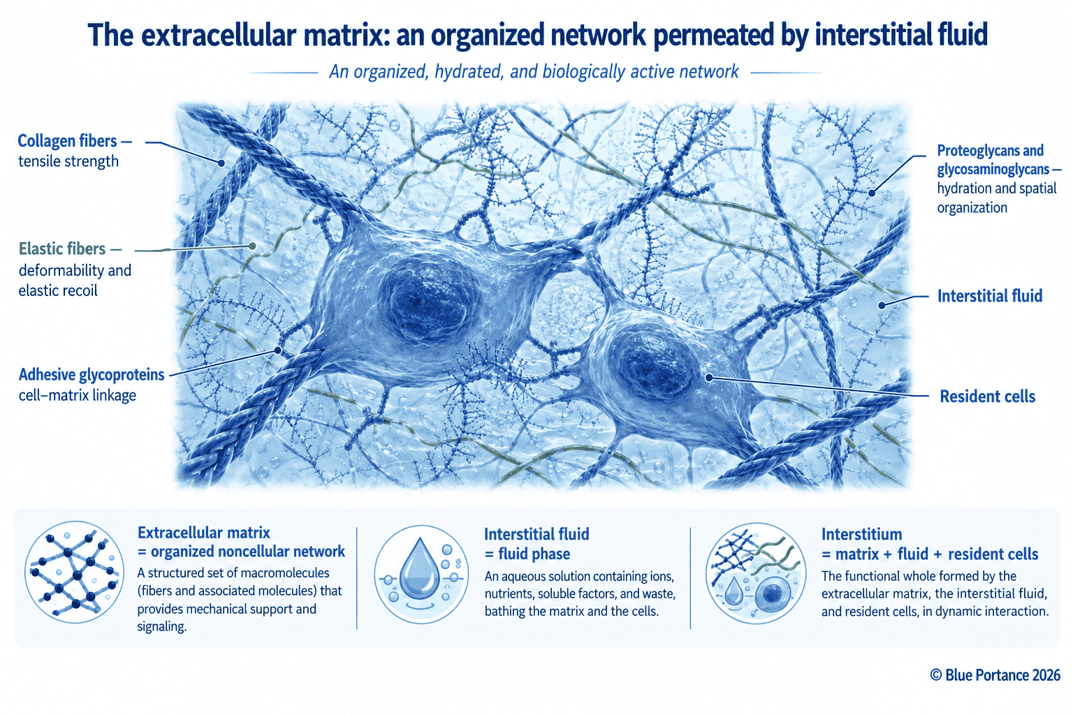
The extracellular matrix comprises several main constituents:
- collagen fibers;
- elastic fibers;
- adhesive glycoproteins;
- proteoglycans;
- glycosaminoglycans;
- a water phase associated with the interstitial fluid.
Collagen contributes mainly to tensile strength, whereas elastic fibers contribute to deformability and elastic recoil. Proteoglycans and glycosaminoglycans, for their part, influence hydration and osmotic properties. They also help organize the extracellular space (Theocharis et al., 2016).
Matrix composition varies considerably from one tissue to another. For example, the matrix of a tendon does not have the same architecture as that of cartilage, muscle or nervous tissue. These differences therefore partly determine how forces are transmitted. They also influence the local movement of water and solutes.
This matrix should therefore be understood as a hierarchical organization. Indeed, its constituents assemble at different scales, from molecular interactions up to the overall architecture of the tissue.
2. A Mechanical Structure, but Not Only That
The extracellular matrix helps maintain the shape of tissues. It also contributes to the distribution of mechanical stresses and to the transmission of forces between cells.
This mechanical function alone, however, does not fully describe its role. It is also involved in:
- cell adhesion;
- orientation;
- migration;
- differentiation;
- survival;
- local storage of certain molecules;
- transmission of mechanical and chemical signals.
The physical and biochemical properties of the matrix can thus influence the behavior of the cells that inhabit it (Hynes, 2009; Yue, 2014).
Nor is the matrix independent of the cells. In fact, the cells produce some of its constituents, organize them, modify them and break them down. They also respond to the composition and mechanical properties of their extracellular environment.
There is thus a reciprocal dialogue between cells and the matrix. This relationship distinguishes the living matrix from an inert material. In other words, it can transmit forces, but it can also be remodeled and interpreted by the cells.
3. Water Associated with Matrix Macromolecules
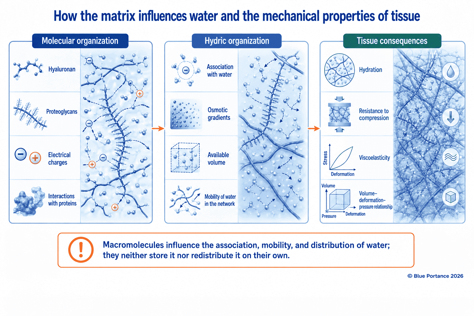
Part of the water present in the extracellular environment interacts with matrix macromolecules. In addition, the nature and organization of these macromolecules influence water retention, water mobility and the mechanical properties of the tissue.
Glycosaminoglycans, and hyaluronan in particular, have hydrophilic properties. Accordingly, they contribute to the hydration and viscoelastic properties of many tissues. Hyaluronan thus helps create a hydrated environment around cells. It is also involved in organizing the matrix (Cowman, 2017).
This capacity to associate with water depends in particular on:
- the type of glycosaminoglycans;
- concentration;
- molecular weight;
- charge;
- interactions with proteins;
- the density of the matrix network.
At the tissue scale, osmotic gradients and the porosity of the network also play a role. Similarly, mechanical stresses influence the distribution and mobility of fluids.
However, matrix macromolecules do not, on their own, “store” or “redistribute” water. Rather, they influence its association, mobility and distribution within the interstitial environment.
A mechanical change in the tissue can thus alter the available volume and the deformation of the network. It can also alter fluid movement, without automatically changing the chemical properties of glycosaminoglycans.
4. The Matrix as a Transport Environment
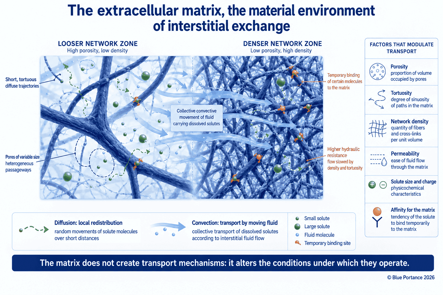
Certain molecules move through the extracellular matrix between blood vessels, cells and the various tissue compartments.
The matrix can:
- offer variable resistance to the movement of molecules;
- temporarily retain certain substances through specific interactions;
- alter local diffusivity depending on the size and charge of solutes;
- influence the paths taken and the distances traveled;
- locally organize certain growth factors and extracellular signals.
The structure of the network, its porosity, its tortuosity and its density all contribute to these phenomena. A molecule may therefore move differently depending on its molecular weight and electric charge. Its affinity for certain components and the organization of the tissue also play a role (Fan et al., 2014).
The matrix does not itself produce diffusion or convection. Instead, it alters the conditions under which they occur by offering hydraulic resistance to fluid movement. It also influences local permeability and imposes a variable hindrance on solutes. Bulk fluid movement can thus carry certain solutes along, while their local redistribution also depends on diffusion. Moreover, the relative contribution of these mechanisms varies with the structure of the matrix, the properties of the solutes and the gradients present.
In other words, the matrix does not systematically “promote” or “block” diffusion. Rather, it alters the conditions under which transfers take place.
This distinction sets the stage for the next chapter of this guide, devoted to the physical mechanisms of diffusion, osmosis, filtration and convection. In contrast, the present chapter focuses primarily on the material medium in which these mechanisms take place.
5. A Time-Dependent Mechanical Organization
The extracellular matrix contributes to the mechanical response of tissues. Depending on its composition and degree of hydration, a tissue can transmit, dissipate or temporarily store part of the mechanical energy it receives.
Tissues also exhibit time-dependent responses. For example, a rapidly applied load, a sustained load and a repeated load do not necessarily produce the same deformation.
Viscoelasticity refers to a response that depends on both deformation and time. Poroelasticity, on the other hand, describes the behavior of a biphasic medium made up of a solid phase and a fluid phase. The relative movements of these two phases contribute to the mechanical response.
In fact, many soft tissues exhibit viscoelastic and poroelastic behavior simultaneously (Elosegui-Artola, 2021; Sowinski et al., 2021).
Depending on loading conditions, tissues can:
- transmit part of the forces;
- spread them over a larger area;
- dissipate part of the energy;
- temporarily store part of the mechanical energy;
- fully or partially recover their initial configuration after unloading;
- retain a deformation when the load is high or prolonged.
However, these properties are not the same in all tissues. In particular, they depend on the composition of the matrix, the amount of fluid and permeability. Fiber architecture and the duration of loading also come into play.
6. Matrix, Interstitial Volume and Pressure
The extracellular matrix does not, on its own, determine interstitial pressure. Through its composition and compliance, however, it contributes to the relationship between interstitial volume and the pressure within it.
Interstitial pressure also depends on:
- microvascular exchange;
- filtration;
- lymphatic uptake;
- the volume of fluid present;
- the properties of the matrix;
- the forces acting within the tissue.
Specifically, Reed and Rubin describe the combined role of interstitial pressure and the extracellular matrix in transcapillary exchange. They also highlight their importance in regulating the interstitial compartment (Reed & Rubin, 2010).
In other words, the extracellular matrix neither produces nor controls interstitial pressure on its own. Instead, it contributes to the mechanical conditions that link interstitial fluid volume, tissue deformation and interstitial pressure.
This relationship will be particularly important for understanding the effects of prolonged compression. It does not mean, however, that a local mechanical stress automatically blocks exchange.
7. An Interface for Cell Signaling
The extracellular matrix does more than provide physical support. It also contributes to the transmission of signals that influence cellular activity.
For example, cells have receptors capable of interacting with certain matrix molecules. These interactions can alter:
- cell shape;
- cytoskeletal organization;
- adhesion;
- migration;
- proliferation;
- differentiation;
- certain signaling pathways.
The stiffness and tension of the matrix can thus influence the cellular response. Its topography and chemical composition play this role as well (Humphrey et al., 2014; Franchi et al., 2024).
These phenomena are among the foundations of mechanobiology and mechanotransduction. They will be developed in a later chapter of this guide.
For now, it is enough to remember that the matrix acts as an interface between the cell and its environment. It conveys mechanical and chemical properties that can alter cellular activity, without functioning as an autonomous nervous or circulatory system.
8. A Continuously Remodeled Matrix
The extracellular matrix is not fixed. Instead, it is produced, organized, modified and broken down according to the needs of the tissue and its physiological state.
This remodeling occurs in particular:
- during development;
- during growth;
- during tissue repair;
- in response to mechanical stresses;
- in inflammatory processes;
- during aging.
For instance, cells can alter the density, orientation and composition of the matrix network. Enzymes can also break down certain components, while other molecules are produced or reorganized.
Moreover, the rate of remodeling varies with the tissues and components involved. An immediate mechanical deformation must therefore be distinguished from longer-term biological remodeling.
Thus, a load applied to a tissue does not instantly transform the matrix. It can, however, alter mechanical signals, cell shape and the local conditions that subsequently contribute to adaptation or remodeling.
9. What This Means for Sitting
Sitting brings the body, the soft tissues and a support surface into contact. This relationship locally alters tissue shape, the distribution of mechanical stresses and the pressure conditions within the tissues and the interstitium.
A support surface does not meet a homogeneous material. Instead, it loads a set of tissues with different compositions, thicknesses and mechanical properties. For example, studies by Makhsous show that seat configuration and certain experimental pressure-relief maneuvers can alter interface pressures. They also show a change in the perfusion measured in the gluteal tissues (Makhsous et al., 2007, 2012). This work thus documents the local consequences of specific mechanical sitting conditions. It does not, however, make it possible to isolate the specific role of the extracellular matrix, nor to conclude that dynamic sitting is clinically effective in general.
Prolonged mechanical stress can locally alter tissue deformation, interstitial volume, pressure and transfer conditions. These effects depend in particular on:
- load intensity;
- duration;
- contact area;
- pressure distribution;
- shear;
- individual tissue properties;
- the possible changes in support configuration.
This situation should not, however, be described as an automatic blockage of the matrix or of exchange. Indeed, tissues remain heterogeneous media, subject to various gradients and to time-dependent responses.
10. Key Takeaways
The extracellular matrix is at once:
- a structural network;
- a mechanical interface;
- a medium hydrated by interstitial fluid;
- a space for local transport;
- a substrate for adhesion;
- a reservoir of extracellular signals;
- an environment for communication with cells.
The extracellular matrix, then, does not simply move fluids around. Rather, it alters the physical and chemical conditions under which water, solutes and signals move.
This framing avoids two opposite oversimplifications. The first treats the matrix as an entirely passive scaffold. The second, in contrast, attributes to it an autonomous ability to “unblock,” “drain” or “rehydrate” tissues.
When sitting, the issue is therefore to examine the duration and distribution of mechanical stresses. It is also to consider the possible changes in support configuration, without turning these general mechanisms into a clinical promise about any particular seat.
Article Summary
In short, the extracellular matrix is the non-cellular network in which cells live, adhere, deform and communicate with their environment. It is composed of proteins, glycoproteins, proteoglycans and glycosaminoglycans. In addition, this matrix is permeated by interstitial fluid and associated with water that contributes to the mechanical properties of the tissue.
Moreover, it can offer variable resistance to the movement of molecules and retain certain solutes. It can also alter their diffusivity and contribute to the relationship between interstitial volume, pressure and deformation (Fan et al., 2014; Reed & Rubin, 2010).
Overall, its state depends on its composition, hydration and organization. The mechanical stresses it receives and its remodeling also contribute. A prolonged load can alter local mechanical and fluid conditions, but it should not be presented as a complete blockage of flows.
The next chapter of this guide will detail the physical mechanisms that allow matter to pass through tissues: diffusion, osmosis, filtration and convective transport.
Scientific References
Structure and Composition of the Extracellular Matrix
- Cowman, M. K. (2017). Hyaluronan and hyaluronan fragments. Advances in Carbohydrate Chemistry and Biochemistry, 74, 1–59. https://doi.org/10.1016/bs.accb.2017.10.001
- Hynes, R. O. (2009). The extracellular matrix: not just pretty fibrils. Science, 326(5957), 1216–1219. https://doi.org/10.1126/science.1176009
- Theocharis, A. D., Skandalis, S. S., Gialeli, C., & Karamanos, N. K. (2016). Extracellular matrix structure. Advanced Drug Delivery Reviews, 97, 4–27. https://doi.org/10.1016/j.addr.2015.11.001
- Yue, B. (2014). Biology of the extracellular matrix: an overview. Journal of Glaucoma, 23(8 Suppl 1), S20–S23. https://doi.org/10.1097/IJG.0000000000000108
Tissue Mechanics: Viscoelasticity and Poroelasticity
- Elosegui-Artola, A. (2021). The extracellular matrix viscoelasticity as a regulator of cell and tissue dynamics. Current Opinion in Cell Biology, 72, 10–18. https://doi.org/10.1016/j.ceb.2021.04.002
- Franchi, M., Piperigkou, Z., Mastronikolis, N. S., & Karamanos, N. K. (2024). Extracellular matrix biomechanical roles and adaptation in health and disease. FEBS Journal, 291(3), 430–440. https://doi.org/10.1111/febs.16938
- Humphrey, J. D., Dufresne, E. R., & Schwarz, U. S. (2014). Mechanotransduction and mechanobiology. Nature Reviews Molecular Cell Biology, 15, 802–812. https://doi.org/10.1038/nrm3896
- Sowinski, D. R., McGarry, M. D. J., Van Houten, E. E. W., et al. (2021). Poroelasticity as a model of soft tissue structure: hydraulic permeability reconstruction for magnetic resonance elastography in silico. Frontiers in Physics, 8, 617582. https://doi.org/10.3389/fphy.2020.617582
Interstitial Transport and Sitting
- Fan, D., Creemers, E. E., & Kassiri, Z. (2014). Matrix as an interstitial transport system. Circulation Research, 114(5), 889–902. https://doi.org/10.1161/CIRCRESAHA.114.302335
- Makhsous, M., Priebe, M., Bankard, J., et al. (2007). Measuring tissue perfusion during pressure relief maneuvers: insights into preventing pressure ulcers. The Journal of Spinal Cord Medicine, 30(5), 497–507.
- Makhsous, M., Lin, F., Hanawalt, D., Kruger, S. L., & LaMantia, A. (2012). The effect of chair designs on sitting pressure distribution and tissue perfusion. Human Factors, 54(6), 1066–1074. https://doi.org/10.1177/0018720812457681
- Reed, R. K., & Rubin, K. (2010). Transcapillary exchange: role and importance of the interstitial fluid pressure and the extracellular matrix. Cardiovascular Research, 87(2), 211–217. https://doi.org/10.1093/cvr/cvq143
