


1. From Force to Cellular Response
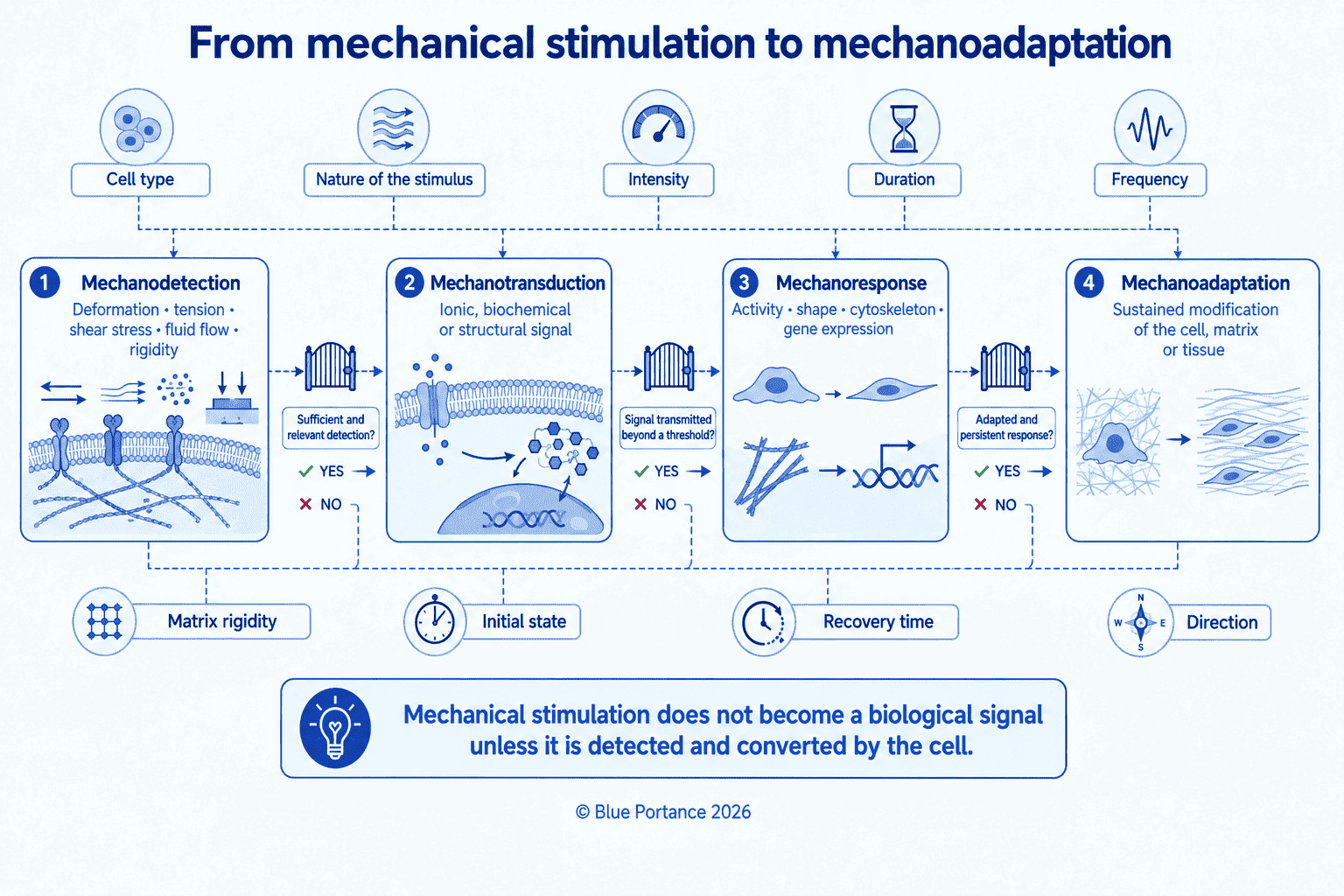
To begin with, several steps need to be distinguished:
- a force is applied to the cell or its environment;
- the cell or the matrix deforms;
- a mechanosensitive structure detects this deformation;
- an intracellular signal is triggered;
- the cell’s activity changes;
- the tissue as a whole may be influenced.
Mechanical force is therefore the initial stimulus, but it is not yet the biological signal. The signal arises when the cell transforms this stress into a molecular or electrical change.
Mechanotransduction is thus a form of conversion between two levels:
- the mechanical level: force, tension, compression, shear, or stiffness;
- the biological level: opening of a channel, activation of an enzyme, change in the cytoskeleton, gene expression, or matrix remodeling.
Cells and tissues indeed have several mechanisms for detecting and interpreting these forces (Wang & Thampatty, 2006; Humphrey et al., 2014).
2. The Structures That Detect Mechanical Stress
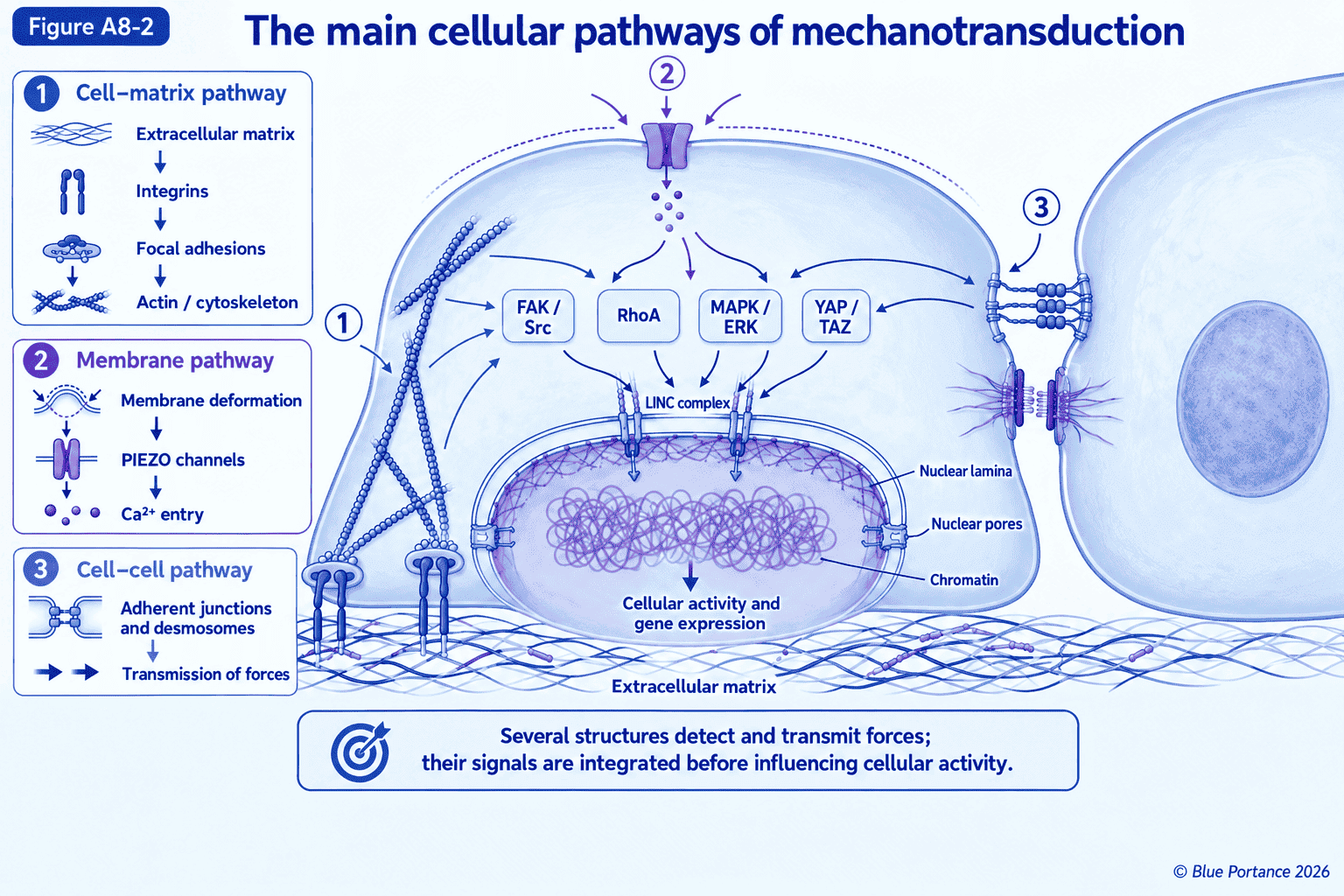
The cell does not have a single mechanical receptor; instead, several structures can take part in detection:
- integrins;
- focal adhesions;
- cell–cell junctions;
- mechanosensitive ion channels;
- the cytoskeleton;
- the nuclear envelope;
- chromatin.
Which structures are engaged depends on how the force is applied and on the function of the cell.
For example, a cell adhering to a stiff matrix does not receive the same mechanical signals as a cell located in a soft matrix, or in an environment subjected to fluid flow. Forces can also be transmitted by neighboring cells or through contacts between cells.
Mechanotransduction should therefore be understood as a detection and transmission network, rather than as a linear chain with a single sensor.
3. Integrins and Focal Adhesions
Integrins are transmembrane proteins that link the extracellular matrix to the cell’s cytoskeleton.
As a result, they establish mechanical continuity between:
- matrix fibers and macromolecules;
- the cell membrane;
- focal adhesion proteins;
- actin filaments;
- the cytoskeleton;
- the interior of the cell.
When a force is exerted on the matrix or on the cell, integrins can transmit that force to intracellular structures. They also take part in activating signaling proteins, particularly at focal adhesions (Ross et al., 2013).
Focal adhesions are therefore not mere anchoring points. Instead, they are mechanochemical platforms, in which a force can alter protein conformation, cytoskeletal organization, and the activity of signaling pathways.
Stress exerted on the matrix can thus alter:
- the tension of actin filaments;
- cell shape;
- adhesion stability;
- migration;
- proliferation;
- extracellular matrix production.
Active Mechanosensing
However, mechanosensing is not purely passive. Indeed, through its contractility, the cell itself pulls on the matrix by means of actomyosin. In return, it then meets a resistance that depends on the organization and stiffness of that matrix. This resistance contributes to adhesion maturation, cytoskeletal tension, and certain signaling pathways. The cell does not merely undergo forces: it also mechanically probes its environment.
4. The Cytoskeleton as a Transmission Network
The cytoskeleton includes, among other components:
- actin filaments;
- microtubules;
- intermediate filaments.
It gives the cell its mechanical organization and also helps carry forces between the membrane, the adhesions, and the nucleus.
However, the cytoskeleton is not a rigid structure. It reorganizes according to the loads it receives, tensions, and chemical signals. In addition, a change in its tension can influence cell shape and the activity of many proteins.
The cytoskeleton therefore acts simultaneously as:
- a transmission system;
- a load-resisting element;
- a regulator of cell shape;
- a relay between the membrane and the nucleus.
Cells can thus respond to the stiffness of their environment by modifying their cytoskeleton and their adhesion. The mechanical properties of the matrix then become biological information that can influence cell behavior (Humphrey et al., 2014).
5. Mechanosensitive Ion Channels
Besides these structures, some membrane proteins can respond to tension or deformation of the membrane. These include, in particular, mechanosensitive ion channels.
When they open, they allow ions such as calcium, sodium, or potassium to pass through. This rapid change in the cell’s electrical and ionic composition can then trigger:
- a change in membrane potential;
- enzyme activation;
- cytoskeletal reorganization;
- release of signaling molecules;
- changes in the expression of certain genes.
Specifically, among mechanosensitive channels, PIEZO1 and PIEZO2 are major examples. Their opening in response to stresses transmitted to the membrane allows cations, particularly calcium, to enter the cell. As a result, this provides a rapid conversion of the mechanical stimulus into an electrochemical signal. Their functions nevertheless differ across cells and tissues. Their presence therefore does not allow one to predict a uniform response to every deformation (Xiao, 2024).
Mechanosensitive channels thus provide a particularly fast conversion pathway between a physical deformation and a cellular response. They are involved in many tissues, but their precise role depends on the cell type, the location of the channel, and the intensity of the stimulation.
One should nonetheless avoid claiming that any pressure applied to a tissue automatically opens these channels. The response depends on the deformation actually transmitted to the membrane and on the cell’s own properties.
6. Cell–Cell Junctions
Furthermore, forces are not exerted only between a cell and the matrix. They can also be transmitted between neighboring cells.
Adherens junctions and desmosomes link cells to one another and contribute to the mechanical continuity of tissues. A pull exerted on one cell can therefore be transmitted to its neighbors.
This transmission allows the tissue to behave as a coordinated organization, and it can also alter intracellular signals in several cells at once.
Cell junctions thus contribute to several functions:
- stress distribution;
- tissue cohesion;
- coordination of responses;
- transmission of mechanical signals;
- collective adaptation of cells.
Mechanotransduction mechanisms can therefore be individual, but they can also be organized at the scale of a tissue.
7. The Nucleus as a Mechanical Integrator
The nucleus is not simply a reservoir of DNA isolated from the rest of the cell. Instead, it is mechanically connected to the cytoskeleton by the LINC complex (Linker of Nucleoskeleton and Cytoskeleton).
Specifically, this complex connects, among other elements:
- cytoskeletal filaments;
- nesprin proteins;
- SUN proteins;
- the nuclear lamina;
- the nuclear envelope.
A force applied to the matrix can therefore be transmitted all the way to the nucleus, through physical continuity between the extracellular environment and the nucleoplasm (Kirby & Lammerding, 2018).
Deformation of the nuclear envelope can then alter:
- the shape of the nucleus;
- nuclear membrane tension;
- the organization of the lamina;
- the function of nuclear pores;
- the arrangement of chromatin.
The nucleus can thus act as a mechanical integrator. It does not detect force consciously; rather, it transforms the deformations it receives into changes in its structure and function.
8. From Mechanosensitive Structures to Signaling Pathways
Between mechanical detection and gene expression, several biochemical pathways come into play. They link mechanosensitive structures to the regulators that alter cellular activity.
In focal adhesions, for example, mechanical stress can activate the kinases FAK and Src. This activation then contributes to the recruitment of signaling proteins and to adhesion maturation.
In addition, the small GTPase RhoA regulates actomyosin contractility and cytoskeletal tension. Its activation can therefore strengthen adhesions and change the shape of the cell.
Calcium entry, particularly through PIEZO channels, can activate calcium-dependent enzymes. It thus contributes to several of these pathways.
The MAPK/ERK pathways can also be engaged by mechanical stress, and they contribute to the regulation of proliferation and gene activity.
YAP and TAZ: Transcriptional Relays
Finally, among the regulators sensitive to these signals, YAP and TAZ hold a special place. These transcriptional coactivators respond in particular to matrix stiffness, cell shape, and cytoskeletal tension. Depending on whether they are located in the nucleus or the cytoplasm, they can modulate the expression of genes involved in proliferation, differentiation, and matrix remodeling (Panciera et al., 2017).
Overall, these pathways do not operate in isolation. They interact with one another and partly converge on shared regulators, which explains why the same mechanical stress can produce different cellular effects depending on the context.
9. From Mechanical Forces to Gene Expression
In particular, deformation of the nucleus can influence exchanges between the cytoplasm and the nucleoplasm. It can also alter chromatin organization and the accessibility of certain regions of the genome.
A force can thus influence gene expression through several intersecting pathways:
- physical transmission all the way to the nucleus, via the cytoskeleton and the LINC complex;
- activation of biochemical pathways such as FAK/Src, RhoA, or MAPK/ERK;
- ion entry, particularly through PIEZO channels;
- translocation of transcriptional regulators such as YAP and TAZ.
Direct deformation of the nucleus is therefore one mechanotransduction pathway among others, not the only pathway leading to gene expression.
The consequences of these combined pathways may involve, for example:
- protein production;
- extracellular matrix synthesis;
- metabolism;
- proliferation;
- differentiation;
- repair;
- the inflammatory response.
Recent research presents the nucleus as an active component of cellular mechanoadaptation, rather than merely the last link in a transmission chain (Echarri, 2022; Uhler & Shivashankar, 2017).
10. A Response That Depends on Magnitude and Duration
The cellular response also depends on the mechanical dose.
For example, a brief, moderate force may cause a transient response. In contrast, a repeated or sustained force may lead to more lasting remodeling.
Specifically, the key parameters are:
- magnitude;
- duration;
- frequency;
- rate of application;
- direction;
- repetition;
- stiffness of the environment;
- initial state of the cell.
A cell can also gradually adapt to repeated stimulation. However, this adaptation does not necessarily mean that the signal disappears. Instead, it may reflect a change in the response threshold, or a reorganization of the mechanosensitive structures.
The mechanical dose is not limited to magnitude and duration, however. The recovery time between two loading episodes also matters, as does whether the load is continuous or intermittent. The spatial and directional variability of the stresses, as well as the tissue’s prestress and the cell’s mechanical history, can also modify the response.
The same average load can thus produce different biological environments, depending on whether it stays constant or alternates with phases of lighter loading. One should nonetheless avoid claiming that this intermittency necessarily leads to beneficial adaptation.
Excessive Stress
Conversely, excessive stress can provoke an unfavorable response:
- cytoskeletal damage;
- rupture of adhesions;
- disruption of the nuclear envelope;
- inflammation;
- apoptosis;
- abnormal matrix remodeling.
One should therefore avoid labeling a mechanical stress as beneficial or harmful independently of its dose and biological context.
11. From Mechanotransduction to Tissue Adaptation
A cell’s response can also have consequences for the tissue.
For example, a mechanically loaded cell can alter:
- collagen production;
- glycosaminoglycan synthesis;
- release of growth factors;
- matrix organization;
- adhesion to neighboring cells;
- metabolic activity.
When several cells respond in a coordinated way, the matrix and the structure of the tissue can be gradually remodeled.
A Loop Rather Than a Chain
This relationship does not run in one direction only, however. Indeed, the properties of the matrix shape the cell’s mechanical response. That response in turn alters the production and organization of the matrix, which then changes the properties to which the cell is exposed. Cell and matrix are thus part of a loop, rather than a simple chain of consequences.
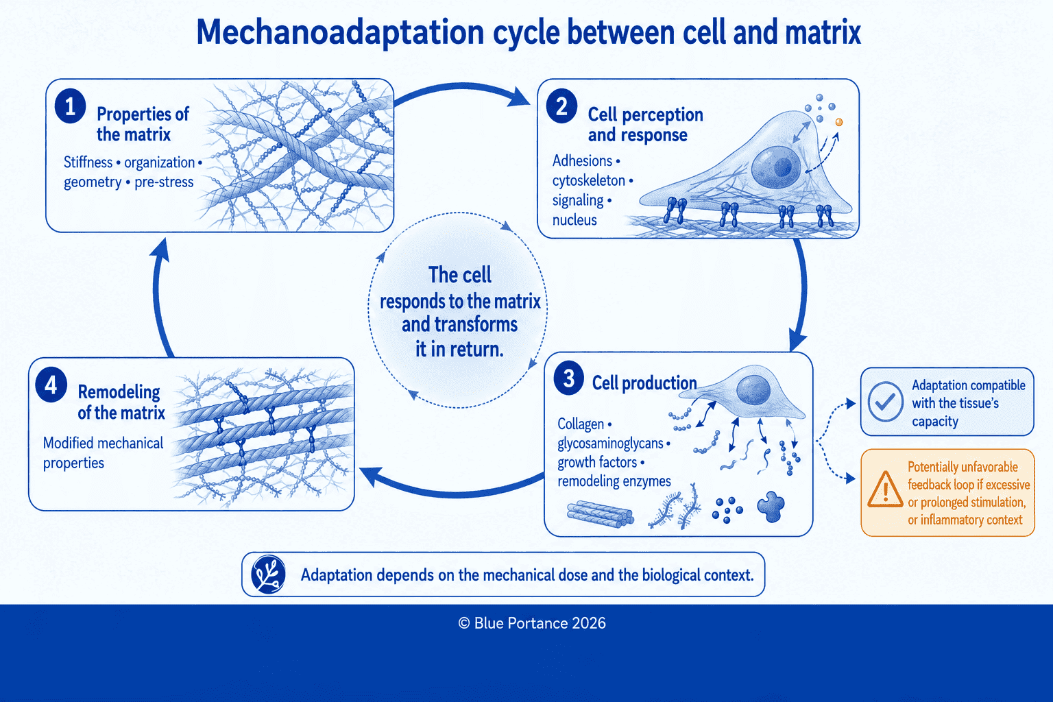
This adaptation can be functional when the stress remains within the tissue’s capacities. It can become unfavorable when the load is excessive, repeated, or associated with inflammation. This type of abnormal loop has also been proposed in certain fibrotic processes.
Mechanotransduction therefore provides a link between:
- macroscopic forces;
- tissue deformation;
- cellular responses;
- biological remodeling.
It does not, however, allow a clinical benefit to be inferred directly from an isolated cellular mechanism.
12. What This Means for Sitting
When we sit, tissues are subjected to compression, deformation, and sometimes shear. As a result, these mechanical demands can alter cell shape, matrix tension, and the relationships between tissues.
A support surface can therefore constitute a mechanical environment for cells. It would, however, be an overstatement to claim that it necessarily triggers a favorable biological response.
The response depends in particular on:
- the magnitude of the stress;
- the area over which it is distributed;
- the duration of the load;
- shear;
- tissue deformability;
- the ability to reposition;
- the biological state of the tissue.
A Spatial and Temporal Environment
From a mechanobiological standpoint, a seat is therefore not characterized solely by an average pressure. Instead, it defines a spatial and temporal mechanical environment that includes, among other factors:
- the spatial distribution of pressures;
- compression gradients through the depth of the tissues;
- shear;
- holding duration;
- repetition of loads;
- the ability to reposition;
- shifting of the most heavily loaded zones over time;
- alternation between loading and lighter loading.
Studies of seating and interface pressures show that support design can change load distribution, as well as certain local perfusion indicators (Makhsous et al., 2007, 2012). However, they do not directly demonstrate that dynamic sitting activates beneficial mechanotransduction or improves cellular remodeling.
In short, a seat modifies the mechanical environment of the tissues. It can thus influence the deformations, stresses, and mechanical signals that cells receive, without this alone making it possible to predict a favorable biological response.
13. Key Takeaways
Mechanical stress becomes a biological signal when it is detected by a mechanosensitive structure and then converted into an intracellular response.
In brief, the main steps are:
- deformation of the matrix or the membrane;
- transmission through integrins and adhesions;
- cytoskeletal reorganization;
- possible opening of ion channels, particularly PIEZO1 and PIEZO2;
- activation of biochemical pathways such as FAK/Src, RhoA, or MAPK/ERK;
- mechanical transmission to the nucleus, or translocation of regulators such as YAP and TAZ;
- changes in nuclear pores or chromatin;
- adaptation of cellular activity.
Above all, mechanotransduction is not an automatic reaction to any pressure. Rather, it is a conversion that depends on the cell type, the structure of the tissue, and the magnitude, duration, and repetition of the stress.
Article Summary
In summary, several systems allow cells to detect mechanical forces. To begin with, integrins link the extracellular matrix to the cytoskeleton. Certain mechanosensitive channels, particularly PIEZO1 and PIEZO2, convert membrane deformations into ionic signals. In addition, junctions transmit forces between cells. Finally, the LINC complex connects the cytoskeleton to the nuclear envelope.
Between these structures and gene expression, biochemical pathways such as FAK/Src, RhoA, or YAP/TAZ carry out the conversion of the signal. Mechanical stress can thus alter cell shape, cytoskeletal tension, nuclear structure, chromatin organization, and the expression of certain genes.
This response nevertheless depends on the mechanical dose and the biological context. Moderate, appropriate stress can contribute to an adaptive response, whereas excessive, repeated, or prolonged loading can contribute to an unfavorable response.
When it comes to sitting, then, the point is not to attribute a specific biological action to every pressure. Instead, it is to understand that the geometry, distribution, duration, and variability of mechanical stresses define the mechanical environment to which cells are exposed.
Scientific References
Mechanotransduction: General Principles
- Humphrey, J. D., Dufresne, E. R., & Schwartz, M. A. (2014). Mechanotransduction and extracellular matrix homeostasis. Nature Reviews Molecular Cell Biology, 15(12), 802–812. https://doi.org/10.1038/nrm3896
- Jaalouk, D. E., & Lammerding, J. (2009). Mechanotransduction gone awry. Nature Reviews Molecular Cell Biology, 10(1), 63–73.
- Wang, J. H.-C., & Thampatty, B. P. (2006). An introductory review of cell mechanobiology. Biomechanics and Modeling in Mechanobiology, 5(1), 1–16.
Adhesions, Cytoskeleton, and Mechanosensitive Channels
- Ross, T. D., Coon, B. G., Yun, S., et al. (2013). Integrins in mechanotransduction. Current Opinion in Cell Biology, 25(5), 613–618. https://doi.org/10.1016/j.ceb.2013.05.006
- Sun, Z., Guo, S. S., & Fässler, R. (2016). Integrin-mediated mechanotransduction. Journal of Cell Biology, 215(4), 445–456.
- Xiao, B. (2024). Mechanisms of mechanotransduction and physiological roles of PIEZO channels. Nature Reviews Molecular Cell Biology, 25(11), 886–903. https://doi.org/10.1038/s41580-024-00773-5
Nucleus, Signaling Pathways, and Remodeling
- Echarri, A. (2022). A multisensory network drives nuclear mechanoadaptation. Biomolecules, 12(3), 404. https://doi.org/10.3390/biom12030404
- Kirby, T. J., & Lammerding, J. (2018). Emerging views of the nucleus as a cellular mechanosensor. Nature Cell Biology, 20(4), 373–381. https://doi.org/10.1038/s41556-018-0038-y
- Panciera, T., Azzolin, L., Cordenonsi, M., & Piccolo, S. (2017). Mechanobiology of YAP and TAZ in physiology and disease. Nature Reviews Molecular Cell Biology, 18(12), 758–770. https://doi.org/10.1038/nrm.2017.87
- Uhler, C., & Shivashankar, G. V. (2017). Regulation of genome organization and gene expression by nuclear mechanotransduction. Nature Reviews Molecular Cell Biology, 18(11), 717–727. https://doi.org/10.1038/nrm.2017.101
Sitting and Interface Pressures
- Makhsous, M., Priebe, M., Bankard, J., et al. (2007). Measuring tissue perfusion during pressure relief maneuvers: insights into preventing pressure ulcers. The Journal of Spinal Cord Medicine, 30(5), 497–507.
- Makhsous, M., Lin, F., Hanawalt, D., Kruger, S. L., & LaMantia, A. (2012). The effect of chair designs on sitting pressure distribution and tissue perfusion. Human Factors, 54(6), 1066–1074. https://doi.org/10.1177/0018720812457681
