


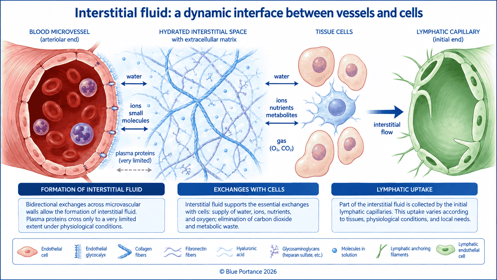
1. Interstitial fluid: a constantly renewed compartment
This fluid is produced mainly through exchange between the microvessels and the extracellular space. Part of the fluid crosses the microvascular wall and enters the interstitium. There, it then comes into contact with the extracellular matrix and the cells.
This fluid contains, among other things:
- water;
- ions;
- nutrients;
- dissolved oxygen and carbon dioxide;
- proteins;
- signaling molecules;
- byproducts of cellular metabolism.
The interstitium therefore comprises a fluid phase, an extracellular matrix, solutes and resident cells. Accordingly, changes in this microenvironment can influence how cells function and how local exchange takes place (Wiig & Swartz, 2012).
Interstitial fluid is not, however, a uniform volume. Indeed, its composition and mobility can vary from one tissue to another. The tissue’s metabolic activity, vascularization, extracellular matrix and uptake pathways also play a role.
2. Formation and uptake
Interstitial fluid is formed mainly through transcapillary exchange. In particular, this exchange is influenced by hydrostatic and oncotic pressures. The permeability of the vessel wall and the properties of the interstitium also play a role.
In many tissues, a small net filtration persists at steady state. As a result, excess interstitial fluid and extravasated macromolecules are collected mainly by the lymphatic vessels. Capillary reabsorption can nevertheless occur transiently, or in certain specialized vascular beds (Levick & Michel, 2010; Wiig & Swartz, 2012).
Lymphatic uptake does not mean that the lymphatic system simply “sucks up” interstitial fluid. Instead, it depends on pressure differences, on the permeability of the initial lymphatics, and on the structure of the endothelial junctions. Further along the network, it also depends on the contractile activity of the collecting vessels (Negrini & Moriondo, 2011; Scallan et al., 2016).
Interstitial renewal therefore results from a balance between:
- fluid formation;
- exchange with cells;
- movement through the matrix;
- uptake by the lymphatics;
- subsequent return to the general circulation.
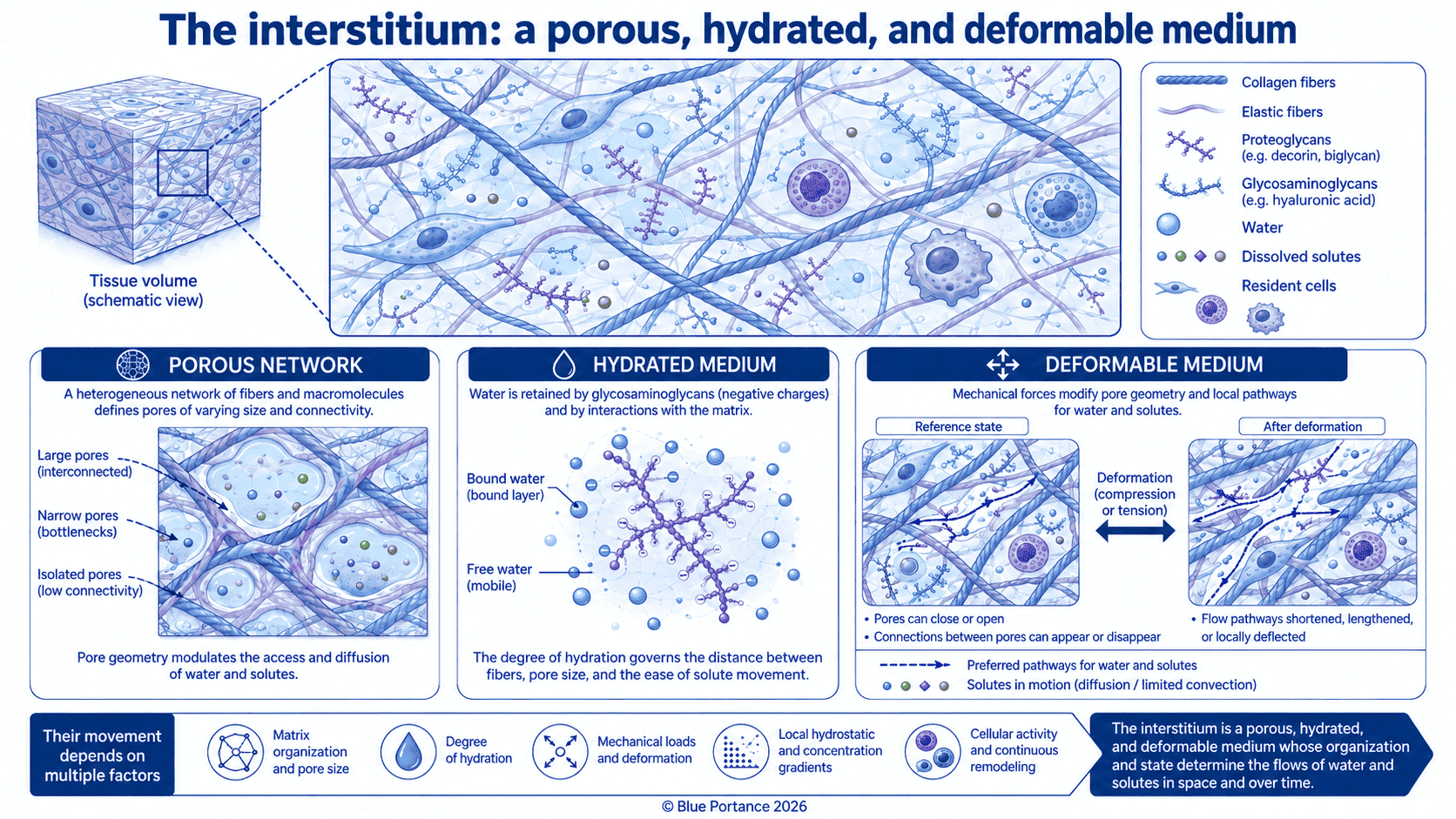
3. The interstitium as a porous, hydrated medium
Interstitial fluid moves through an environment that is nothing like an empty pipe. Indeed, the extracellular matrix forms a complex network of fibers, macromolecules and hydrated spaces.
Fluid mobility depends in particular on:
- the porosity of the network;
- tortuosity;
- fiber density;
- the amount of water bound to macromolecules;
- local permeability;
- interstitial pressure;
- interactions between the fluid and the matrix.
The matrix can therefore offer varying resistance to the movement of fluid and solutes. It can also temporarily retain certain molecules or alter their local distribution (Fan et al., 2014).
In turn, the movement of interstitial fluid can change the mechanical properties of the tissue. There is thus a coupling between the fluid phase and the solid phase of the matrix.
4. Pressure gradients
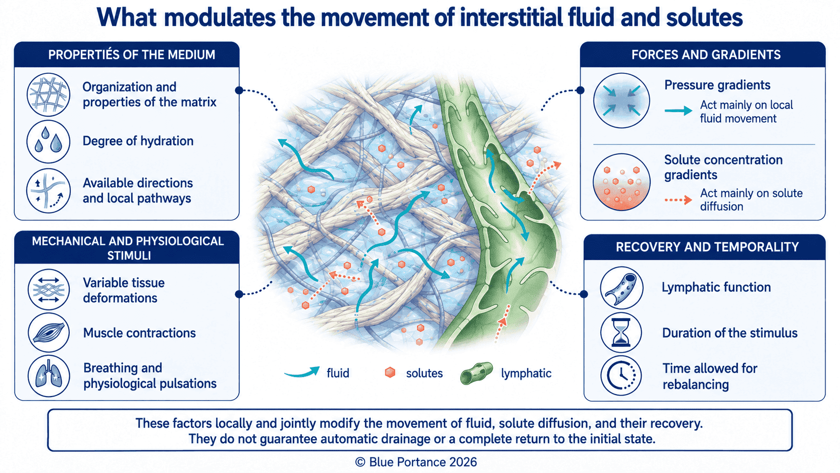
Interstitial fluid moves when differences in pressure or potential exist between different regions of the tissue.
These gradients can be created or altered, for example, by:
- microvascular filtration;
- pressure changes in the capillaries;
- collection by the initial lymphatics, which may help maintain local pressure and flow gradients;
- tissue deformation;
- muscle contraction;
- breathing movements;
- vascular pulsations;
- external compression.
Interstitial pressure does not depend solely on the amount of fluid present. It also depends on the compliance of the interstitium and on the properties of the matrix. Finally, whether the fluid is able to move or be taken up matters as well.
For example, Reed and Rubin show that interstitial pressure and the properties of the extracellular matrix take part in transcapillary exchange. They also highlight their role in regulating the interstitial compartment (Reed & Rubin, 2010).
However, not every external pressure should be equated with a drainage mechanism. Of course, pressure applied to a tissue can displace fluid locally. Its effect, though, depends on its duration, its area and its direction. The deformability of the tissue and the pathways available for redistribution also matter.
5. Compression and decompression
When a tissue is compressed, its volume, shape and fluid distribution can change. As a result, some of the fluid may move toward neighboring regions, or toward spaces where pressure is lower.
Conversely, during decompression, tissues may fully or partially recover their previous configuration. The fluid can then redistribute through the extracellular network.
Still, these phenomena are not instantaneous, nor are they necessarily fully reversible. Indeed, the response depends on:
- loading rate;
- duration of compression;
- cycle frequency;
- tissue structure;
- water content;
- matrix permeability;
- the condition of the blood vessels and lymphatics.
For example, a poroelastic model of the interaction between interstitial fluid and matrix under confined indentation shows that loading and unloading phases can be accompanied by fluid movements in opposite directions. It also indicates that pressure changes are especially large beneath the area where the load is applied (Lu & Wang, 2008).
These findings support the idea of a coupling between solid deformation and fluid movement. However, they do not support the conclusion that there is uniform circulation or systematic drainage.
6. The role of tissue movement
Tissue movement can change local volumes, pressures and gradients. Several mechanical sources may be involved, such as:
- muscle contractions;
- breathing movements;
- arterial pulsations;
- changes in tissue tension;
- organ movement;
- changes in support;
- deformation related to everyday activity.
Initial lymphatics do not have the same contractile apparatus as collecting lymphatic vessels. They rely in particular on the movements of, and the mechanical stresses exerted by, the surrounding tissues.
For instance, Negrini and Moriondo indicate that pressure gradients between the interstitium and the initial lymphatics can be influenced by tissue movement and muscle activity. Heartbeats and breathing also play a role (Negrini & Moriondo, 2011).
In collecting lymphatic vessels, propulsion also depends on intrinsic contractions of the vessel wall and on valve function. Lymph movement therefore results from a combination of extrinsic and intrinsic forces (Scallan et al., 2016).
Breathing movements, for example, are one of these extrinsic forces. During spontaneous inspiration, the descent of the diaphragm lowers intrathoracic pressure, while abdominal pressure generally rises. As a result, these cyclic changes can alter pressure gradients regionally. They may thus contribute to the forward movement of lymph, together with the collecting vessels’ own contractions and the function of their valves. Their effect, however, depends on the region in question, on posture, on breathing depth and on the mechanical properties of the surrounding tissues (Negrini & Moriondo, 2011; Scallan et al., 2016).
7. The specific role of muscle contraction
Muscle contraction should not be confused with simple passive displacement of tissue.
In fact, contractions can actively change local geometry, compress certain spaces and create pressure changes. Consequently, these changes may influence fluid movement and lymph formation.
Similarly, research on lymphatic biomechanics shows that lymphatics located in muscle tissue can undergo cycles of compression and expansion. Specifically, these cycles are linked to the mechanical stresses exerted by the surrounding tissue (Negrini & Moriondo, 2011).
However, how effective this mechanism is depends on the type of movement, its amplitude and its duration. The location of the tissue and the function of the lymphatic vessels also matter. It is therefore not enough to claim that any movement automatically “pumps” interstitial fluid.
In short, muscle contractions can help change tissue pressures and support certain mechanisms of fluid movement and uptake.
8. Interstitial mobility and cellular exchange
The movement of interstitial fluid can contribute to the local transport of nutrients and to the removal of certain metabolic byproducts. It can also change the conditions under which cells receive chemical or mechanical signals.
In avascular tissues, such as adult articular cartilage, exchange with cells relies in particular on solute transport by diffusion and by convection through the extracellular matrix. Moreover, the relative contribution of these mechanisms depends on the properties of the solute and the matrix, and on mechanical loading (Yao & Gu, 2007).
Convection carries solutes along with the bulk movement of fluid, while diffusion contributes to their redistribution along their concentration gradients. These two mechanisms can therefore act simultaneously without being equivalent.
Interstitial fluid should therefore not be viewed as mere waste awaiting removal. Instead, it is a functional compartment in which exchange takes place between the matrix, the cells and the transport networks.
9. What this means for sitting
Sitting places mechanical stresses on soft tissues and changes their shape locally. In particular, prolonged loading can influence:
- tissue deformation;
- interstitial pressure;
- local fluid distribution;
- perfusion conditions;
- the potential for fluid uptake;
- interface pressures.
For example, studies by Makhsous show that seat design and certain experimental pressure-relief maneuvers can change pressure distribution. They also show a change in perfusion measured in the gluteal tissues (Makhsous et al., 2007, 2012). In other words, these studies document the local consequences of specific mechanical configurations. However, they do not make it possible to isolate the specific role of interstitial fluid, the extracellular matrix, or spontaneous micro-movements.
Sustained sitting can therefore reduce changes in mechanical configuration in a given region. It is nevertheless better to avoid the phrase “fluid stagnation,” which suggests a complete halt in exchange.
Load sustained over time, combined with limited opportunities for repositioning, can alter local pressures and deformation. Moreover, the conditions for interstitial fluid renewal depend on these as well.
10. Key takeaways
Interstitial fluid is a dynamic compartment. It is formed by microvascular exchange and then moves through a hydrated matrix network. In addition, it exchanges with cells and is gradually taken up by the lymphatics.
Furthermore, its mobility depends on:
- pressure gradients;
- filtration;
- the extracellular matrix;
- tissue deformation;
- muscle contractions;
- breathing;
- vascular pulsations;
- lymphatic function.
Movement is not the sole driver of interstitial exchange. It can nevertheless change pressures, volumes and the local conditions for fluid movement.
Thus, this conclusion helps avoid two errors: speaking of automatic mechanical drainage, or assuming that immobility immediately shuts down all exchange.
Article summary
Overall, interstitial fluid is a mobile interface between the microvessels, the extracellular matrix and the cells. In other words, it is neither an empty space nor a static reservoir.
Its formation depends on transcapillary exchange, while its mobility depends on gradients, on the properties of the matrix network and on tissue deformation. Finally, its uptake involves the lymphatics, whose function combines intrinsic and extrinsic forces.
Muscle contractions, breathing, vascular pulsations and certain deformations can help change the local conditions for fluid movement and uptake. These effects nonetheless remain dependent on the tissue, the load and the mechanical context.
When sitting, then, the goal is not to indiscriminately “get all bodily fluids circulating again.” Rather, it is to examine how the duration of loading and tissue deformation influence local pressures. Opportunities for changes in support also matter for interstitial renewal.
Scientific References
Interstitium, matrix and tissue transport
- Fan, D., Creemers, E. E., & Kassiri, Z. (2014). Matrix as an interstitial transport system. Circulation Research, 114(5), 889–902. https://doi.org/10.1161/CIRCRESAHA.114.302335
- Wiig, H., & Swartz, M. A. (2012). Interstitial fluid and lymph formation and transport: physiological regulation and roles in inflammation and cancer. Physiological Reviews, 92(3), 1005–1060. https://doi.org/10.1152/physrev.00037.2011
- Yao, H., & Gu, W. Y. (2007). Convection and diffusion in charged hydrated soft tissues: a mixture theory approach. Biomechanics and Modeling in Mechanobiology, 6(1–2), 63–72. https://doi.org/10.1007/s10237-006-0040-3
Interstitial pressure and microvascular exchange
- Levick, J. R., & Michel, C. C. (2010). Microvascular fluid exchange and the revised Starling principle. Cardiovascular Research, 87(2), 198–210. https://doi.org/10.1093/cvr/cvq062
- Lu, Y., & Wang, W. (2008). Interaction between the interstitial fluid and the extracellular matrix in confined indentation. Journal of Biomechanical Engineering, 130(4), 041011. https://doi.org/10.1115/1.2939310
- Reed, R. K., & Rubin, K. (2010). Transcapillary exchange: role and importance of the interstitial fluid pressure and the extracellular matrix. Cardiovascular Research, 87(2), 211–217. https://doi.org/10.1093/cvr/cvq143
Lymphatic function and sitting
- Negrini, D., & Moriondo, A. (2011). Lymphatic anatomy and biomechanics. The Journal of Physiology, 589(Pt 12), 2927–2934. https://doi.org/10.1113/jphysiol.2011.206672
- Scallan, J. P., Zawieja, S. D., Castorena-Gonzalez, J. A., & Davis, M. J. (2016). Lymphatic pumping: mechanics, mechanisms and malfunction. The Journal of Physiology, 594(20), 5749–5768. https://doi.org/10.1113/JP272088
- Makhsous, M., Priebe, M., Bankard, J., et al. (2007). Measuring tissue perfusion during pressure relief maneuvers: insights into preventing pressure ulcers. The Journal of Spinal Cord Medicine, 30(5), 497–507.
- Makhsous, M., Lin, F., Hanawalt, D., Kruger, S. L., & LaMantia, A. (2012). The effect of chair designs on sitting pressure distribution and tissue perfusion. Human Factors, 54(6), 1066–1074. https://doi.org/10.1177/0018720812457681
