


1. The internal environment: a foundational concept
In the 19th century, Claude Bernard formulated a foundational idea. In his view, cells live in a relatively stable internal environment, distinct from the external environment (Bernard, 1865). This concept later transformed physiology profoundly.
The organism does not merely place its cells in contact with the environment. Instead, it actively builds and regulates that environment. Specifically, temperature, pH, ion concentrations and the availability of certain substances are kept within ranges compatible with cellular activity.
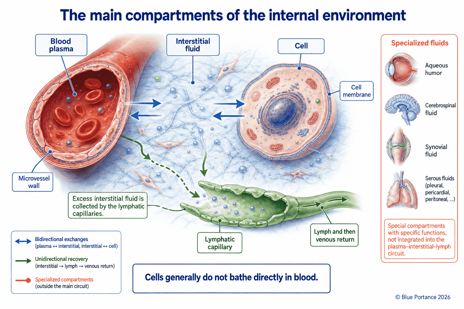
The “internal environment” is therefore not limited to the blood. In fact, it refers more broadly to all the extracellular fluids that surround cells and mediate their exchanges with the rest of the organism. The main compartments involved are the following:
- plasma, located inside the vessels;
- interstitial fluid, present around the cells;
- lymph, derived from interstitial fluid collected by the lymphatic vessels;
- certain specialized fluids, such as cerebrospinal fluid.
In practice, extracellular fluid forms the immediate environment of many cells. In particular, it supplies them with the substances they need and, in return, receives the products of their activity (Boron & Boulpaep, 2017).
2. The extracellular compartments
To begin with, plasma is the liquid fraction located inside the blood vessels. Among other things, it carries water, ions, nutrients, dissolved gases, hormones and proteins.
Part of the water and solutes crosses the microvascular walls. In doing so, it helps form the interstitial fluid that surrounds the cells within tissues. A fraction of this interstitial fluid, along with the macromolecules it contains, is subsequently collected by the lymphatic capillaries. Once it enters the lymphatic network, this fluid is called lymph. It then circulates through a specialized network before returning to the bloodstream (Boron & Boulpaep, 2017; Wiig & Swartz, 2012).
In addition to these compartments, there are specialized fluids such as cerebrospinal fluid, synovial fluid and certain serous fluids. Moreover, their composition and renewal are governed by specific physiological requirements.
These compartments communicate with one another, but they are not interchangeable. Indeed, their compositions, their barriers and their functions differ. As a result, this organization allows the body to maintain local environments suited to the needs of different tissues.
3. In most tissues, cells are not in direct contact with blood
In most tissues, cells are not directly exposed to circulating blood. Plasma remains inside the vessels, while the cells are bathed in interstitial fluid.
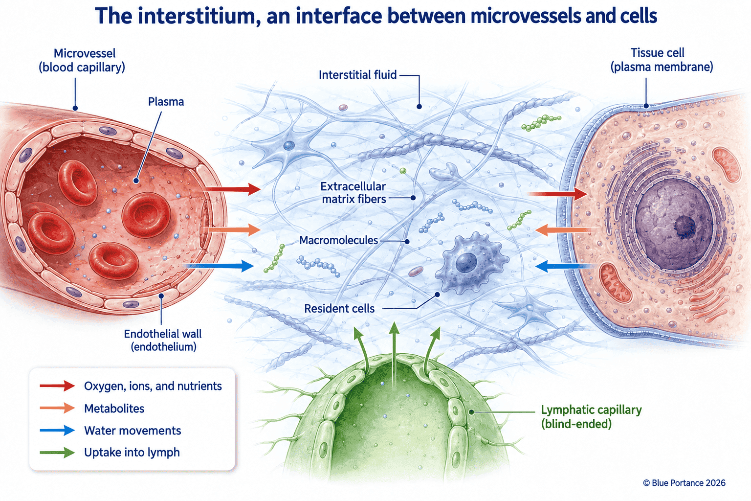
Consequently, exchanges between these two compartments take place across the walls of the microvessels. Interstitial fluid thus serves as an interface between the vascular compartment and the cellular environment.
This organization therefore places several barriers and transfer spaces between plasma and cells. The composition of the cellular environment depends in particular on:
- microvascular permeability;
- the properties of the interstitium;
- the extracellular matrix;
- cellular activity;
- local pressures;
- the uptake of interstitial fluid by the lymphatic system.
Blood thus contributes to maintaining the internal environment. Nevertheless, it is not the direct environment of all cells. This distinction matters because it keeps us from reducing the physiology of exchange to blood circulation alone. Between the blood and the cell, there are in fact interfaces, gradients and local transformations.
4. The interstitium and interstitial fluid
This term refers to the extracellular space located between the cells and the microvessels. Specifically, it includes a fluid phase — the interstitial fluid — an extracellular matrix, solutes and various resident cells. Interstitial fluid is therefore one component of the interstitium, but the two terms are not synonymous.
The interstitium should not be thought of as a mere empty space filled with water. On the contrary, it is an organized microenvironment whose properties depend on several factors:
- the quantity and composition of the fluid;
- the structure of the extracellular matrix;
- the presence of proteins and macromolecules;
- interstitial pressure;
- the permeability of the microvessels;
- the activity of neighboring cells.
For instance, Wiig and Swartz describe the interstitium as a microenvironment made up of fluid, proteins, solutes and extracellular matrix. They also emphasize that changes in it can influence cell function and fluid transport within tissues (Wiig & Swartz, 2012).
Interstitial fluid is thus a dynamic interface between the microvessels, the matrix and the cells.
5. A continuously regulated composition
To function properly, a cell must maintain concentration differences between its interior and its surroundings. These gradients involve, in particular:
- sodium, potassium, calcium and chloride;
- bicarbonate and glucose;
- oxygen and carbon dioxide;
- various proteins and dissolved molecules.
pH and temperature must also remain within ranges compatible with enzyme activity and metabolic reactions.
However, the stability of the internal environment does not mean that these parameters remain perfectly constant. In reality, they fluctuate with physical activity, diet, breathing, temperature and physiological state. Some variations may also be local and depend on the specific activity of a given tissue.
Acid-base balance, for example, is a case of complex regulation. Specifically, extracellular pH depends on buffer systems, ion exchange, respiration and kidney function (Boron & Boulpaep, 2017; Kraut & Madias, 2010).
The internal environment should therefore be understood as a regulated composition. In other words, it is not a uniform fluid with identical properties throughout the body.
6. Distinct tissue microenvironments
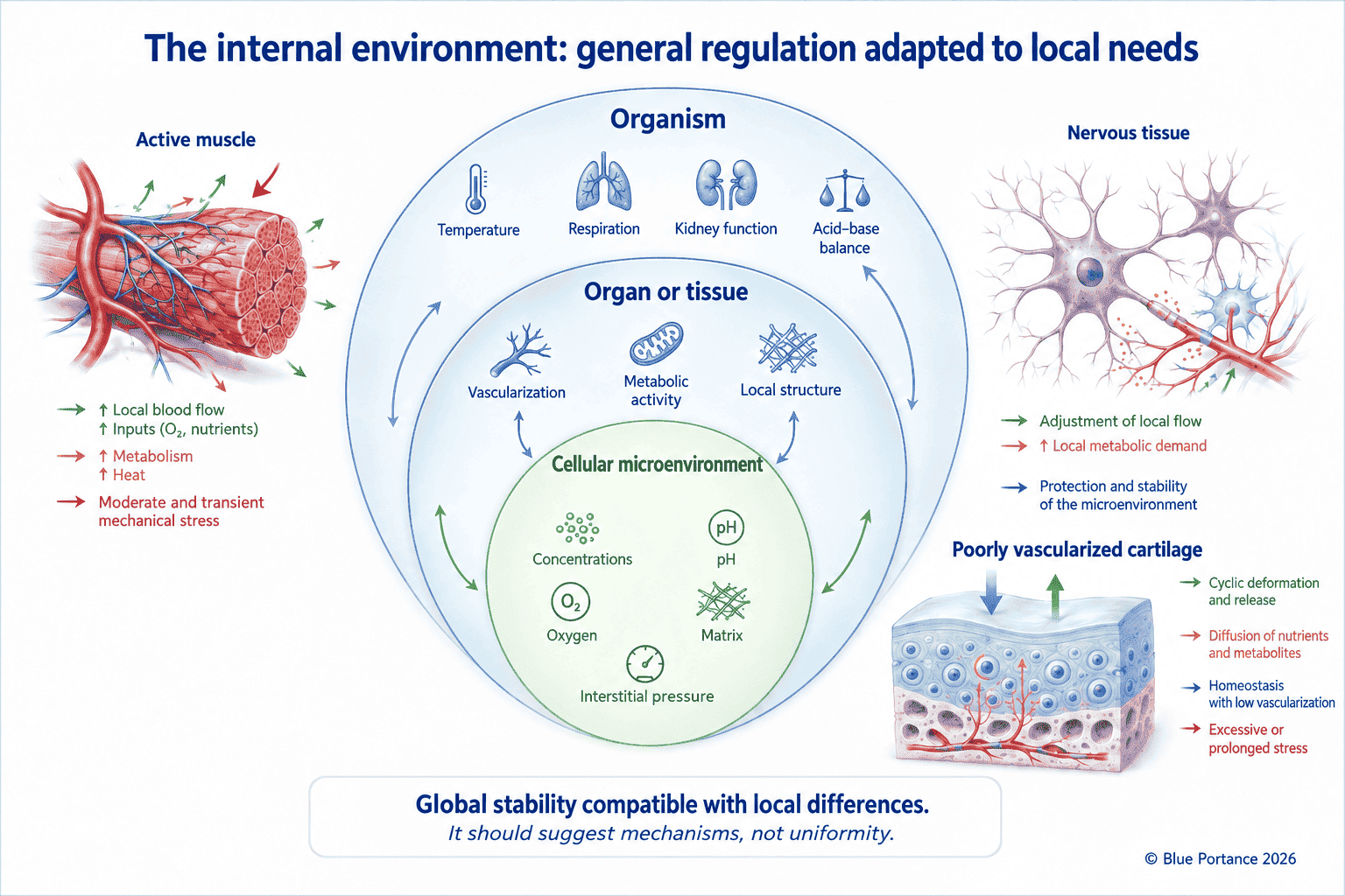
The overall stability of the internal environment does not imply perfect uniformity throughout the body. Indeed, the cells of an active muscle, of nervous tissue, of cartilage or of an epithelium do not experience the same local conditions. Their environment depends on their metabolic activity, their blood supply, the extracellular matrix, temperature and uptake pathways.
For example, a contracting muscle locally increases its oxygen and energy needs. Nervous tissue, in contrast, depends on particularly precise ion gradients. Finally, cartilage, which has a limited blood supply, relies on exchange mechanisms different from those of richly vascularized tissue.
The internal environment should therefore be understood as a nested organization:
- general regulation at the level of the whole organism;
- specific regulation at the level of organs;
- local variations around the cells.
Still, this heterogeneity is not necessarily a flaw. On the contrary, it allows tissues to meet different needs. It does mean, however, that a measurement taken in one compartment cannot automatically be used to infer the state of another.
7. Exchanges among several compartments
Maintaining the internal environment relies on constant exchanges between plasma, the interstitium and cells. These exchanges involve water, ions, nutrients, respiratory gases, hormones, proteins and metabolic products.
They do not, however, rely on a single mechanism. Depending on the substance and tissue involved, they may involve filtration, diffusion, convective transport or membrane transport. These mechanisms will be covered in detail in a later chapter devoted to tissue exchange.
For now, the key point is enough. In short, cells depend on an ongoing balance between the supply of the substances they need and the removal of the products they no longer need. A tissue whose extracellular environment is altered is therefore not necessarily cut off from exchange right away. Its local composition, gradients and transfer conditions, on the other hand, may be altered.
8. Pressures and fluid balance
The movement of water between the microvessels and the interstitium depends on several factors:
- hydrostatic and oncotic pressures;
- the permeability of the vascular wall;
- the properties of the interstitium and the extracellular matrix;
- lymphatic uptake.
Notably, the current understanding of these exchanges has led to a revision of the classic formulation of the Starling principle. Consequently, microvascular exchange can no longer be described as a simple balance between two forces that mechanically cancel each other out. In particular, the endothelial glycocalyx and the local characteristics of the vascular wall play an important role (Levick & Michel, 2010).
In many tissues at steady state, capillaries produce a small net filtration into the interstitium. The excess fluid and macromolecules are then mainly recovered by the lymphatic system. Capillary reabsorption may nevertheless occur transiently, or in certain specialized vascular beds (Levick & Michel, 2010; Reed & Rubin, 2010).
Interstitial pressure and matrix properties also contribute to the conditions of transfer between the microvessels and the tissues (Reed & Rubin, 2010). This point is important. Indeed, the balance of the internal environment does not depend solely on the amount of fluid present. It also depends on the properties of barriers, gradients and uptake pathways.
9. What this means for sitting
Sitting does not bring cells into direct contact with an external object. It first alters the mechanical relationships between the body, the tissues and the support surface. For example, when seated, seat configuration and pressure-relief maneuvers can alter the distribution of interface pressures. They also influence measured perfusion in the gluteal tissues. These effects depend in particular on the magnitude and duration of the load, the contact area, tissue properties and the opportunities for repositioning (Makhsous et al., 2007, 2012). More broadly, prolonged mechanical loading can locally alter interstitial pressure and the conditions under which tissue exchange takes place (Reed & Rubin, 2010; Makhsous et al., 2007, 2012).
This does not mean, however, that a compressed area immediately becomes a closed space, or that exchange stops there entirely. The consequences instead depend on many factors:
- the magnitude of the load and the duration of exposure;
- the contact area and the type of tissue;
- the ability to reposition;
- vascular and metabolic status;
- individual sensitivity.
This article therefore does not yet allow us to conclude that dynamic sitting improves exchange. In fact, it only helps explain why the local environment of cells depends on regulated mechanical, fluid and biological conditions. The following articles in this guide will cover the extracellular matrix, and then the physical mechanisms — diffusion, osmosis, filtration — that make these exchanges possible.
Scientific References
Internal environment and compartments
- Bernard, C. (1865). Introduction à l’étude de la médecine expérimentale. Paris: J.-B. Baillière.
- Boron, W. F., & Boulpaep, E. L. (2017). Medical Physiology (3rd ed.). Philadelphia: Elsevier.
- Kraut, J. A., & Madias, N. E. (2010). Metabolic acidosis: pathophysiology, diagnosis and management. Nature Reviews Nephrology, 6(5), 274–285.
- Wiig, H., & Swartz, M. A. (2012). Interstitial fluid and lymph formation and transport: physiological regulation and roles in inflammation and cancer. Physiological Reviews, 92(3), 1005–1060.
Microvascular exchange and sitting
- Levick, J. R., & Michel, C. C. (2010). Microvascular fluid exchange and the revised Starling principle. Cardiovascular Research, 87(2), 198–210.
- Makhsous, M., Priebe, M., Bankard, J., et al. (2007). Measuring tissue perfusion during pressure relief maneuvers: insights into preventing pressure ulcers. The Journal of Spinal Cord Medicine, 30(5), 497–507.
- Makhsous, M., Lin, F., Hanawalt, D., Kruger, S. L., & LaMantia, A. (2012). The effect of chair designs on sitting pressure distribution and tissue perfusion. Human Factors, 54(6), 1066–1074.
- Reed, R. K., & Rubin, K. (2010). Transcapillary exchange: role and importance of the interstitial fluid pressure and the extracellular matrix. Cardiovascular Research, 87(2), 211–217.
