


This article is part of the Expert Guide “Fascia.”
Chapter 8 — Blue Portance Knowledge BaseHow Can Fascia Contribute to Pain?
Summary — A fascia can contribute to pain when mechanical stress, inflammation, or chemical mediators stimulate its nerve endings, alone or in combination. A loss of gliding can also increase the load on neighboring muscles, nerves, or blood vessels. It thus shifts stress from one tissue to another. When the signals persist, peripheral and then central sensitization can amplify the response. This chapter walks through this sequence step by step.
1. A possible nociceptive source
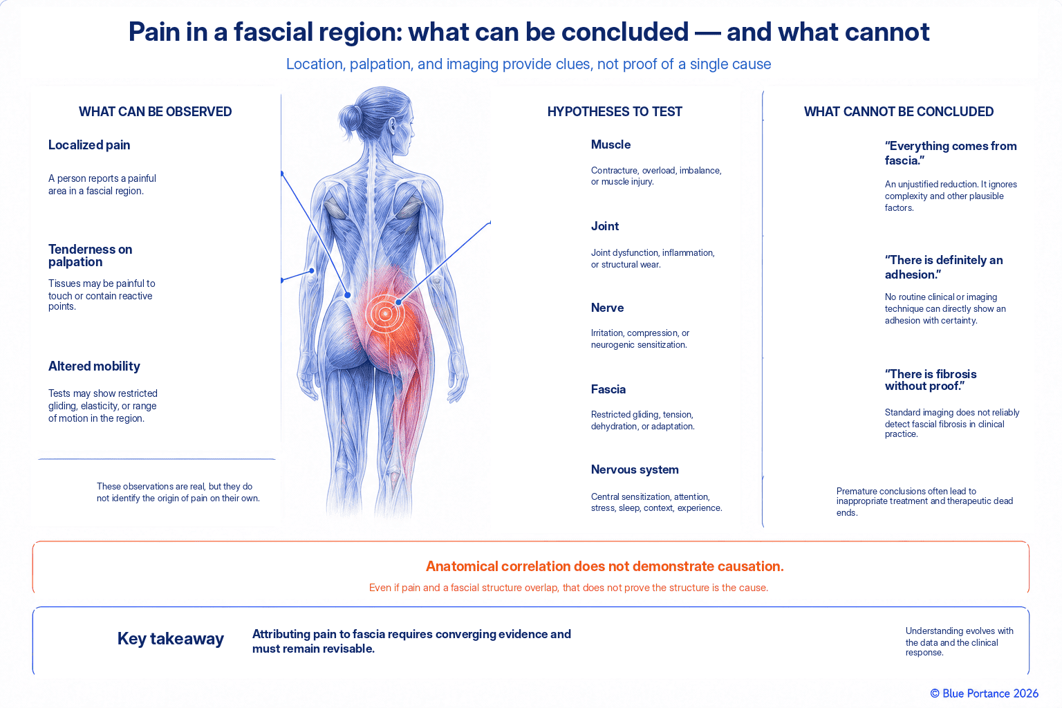
The nerve endings present in certain fasciae can generate nociceptive signals when they receive sufficient stimulation or have become sufficiently sensitized (Suarez-Rodriguez et al., 2022). Pressure, stretching, or inflammation can activate or sensitize free nerve endings. This possibility does not mean that pain without a visible lesion is necessarily fascial. The absence of a lesion visible on imaging rules out neither a fascial origin nor any other tissue origin. This therefore limits the value of imaging alone in settling the question.
A biological framework, not diagnostic certainty
This chapter builds directly on the innervation described in Chapter 7. Some fasciae do contain nociceptive endings, which allows the tissue to contribute, under certain conditions, to a pain experience. This biological possibility should not, however, be turned into diagnostic certainty.
The presence of endings that can be activated by sufficiently intense stimulation is not enough to draw a conclusion. The same holds for weaker stimulation after sensitization. Not all pain felt in a fascia-rich region necessarily comes from this tissue. It may also come from a neighboring structure — muscle, joint, skin, or nerve — that shares the same anatomical area.
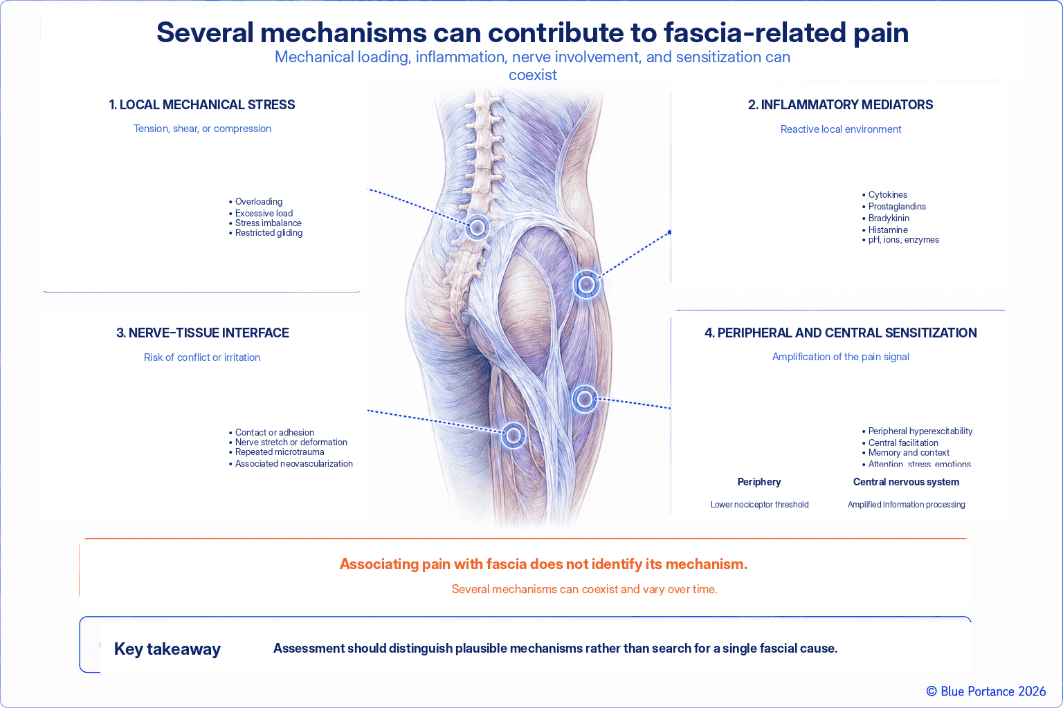
The stimuli capable of activating these endings are themselves varied. Excessive traction, prolonged compression, or the presence of chemical mediators released during inflammation can each cross the activation threshold of a fascial nociceptor. This can happen in isolation or in combination. This plurality of possible triggers makes it all the harder to identify, in retrospect, the precise cause of a given pain episode. The difficulty is especially marked when several factors combine over time. They then no longer act in an isolated, clearly identifiable way.
Three levels of evidence not to be confused
Establishing that a fascia can contribute to pain actually requires clearing several distinct levels of evidence. It is useful to keep them separate. The first level, histological, establishes the anatomical presence of nerve fibers in a given fascia.
The second level, experimental, establishes that controlled stimulation of this tissue can actually produce a measurable nociceptive response. In healthy volunteers, for example, an injection of hypertonic saline into the thoracolumbar fascia caused more intense and longer-lasting local pain. An equivalent injection into the underlying muscle did not produce the same effect (Schilder et al., 2014).
This result demonstrates marked nociceptive sensitivity of this particular fascia under precise experimental conditions. It does not, however, demonstrate that a given episode of clinical low back pain originates in this same tissue. The third level is clinical. It involves attributing actual pain to a specific fascial origin in a given person, which remains the hardest to establish. It requires ruling out or weighing the contribution of other innervated tissues in the same region. No single test can currently do this with certainty.
2. Direct stress and interface stress
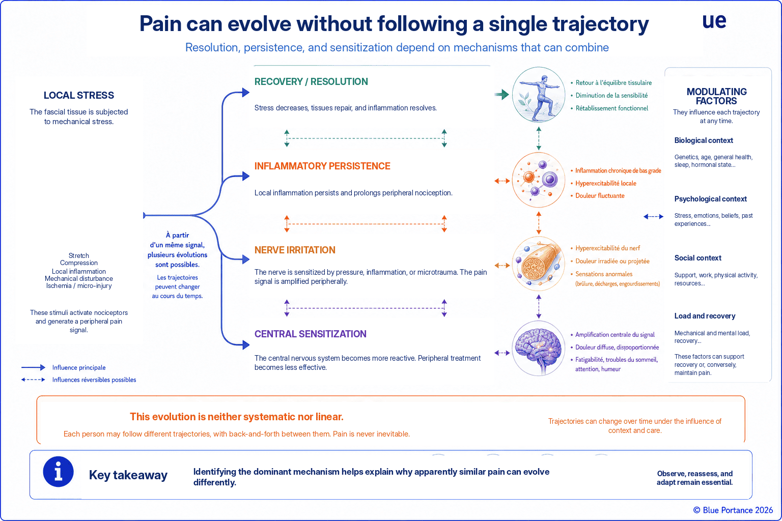
Pain can originate in the tissue itself or in a neighboring structure whose mobility is restricted. A less mobile nerve, an overloaded muscle, or a sensitive scar may coexist. The analysis must therefore cover the entire interface. Chapter 4 showed that mobility between several tissue layers protects each of them by distributing displacements. When the relative mobility between some of these layers decreases or is redistributed, however, this distribution becomes less effective. A normally protected structure may then find itself exposed to stresses it did not encounter before. This type of mechanical redistribution can contribute to symptoms arising from a neighboring structure. This therefore limits the relevance of attributing pain to a single isolated tissue. No study, however, can quantify the share of pain explained by this specific mechanism. Direct injury to the fascia itself could just as well be the cause.
Direct stress and interface stress
To clarify the reasoning, two situations can be distinguished here, and they may combine: a direct nociceptive contribution from the fascia, and an indirect mechanical contribution via a neighboring interface. This distinction is not, however, a recognized pathophysiological classification. It is an analytical teaching tool specific to this chapter. It takes up the distinction made in Chapter 7 between fascia as a direct source of signals and fascia as the mechanical environment of another structure. Direct stress corresponds to stimulation of the fascia’s own nociceptive endings — for example, during excessive stretching or local inflammation.
Three neighboring structures, three mechanisms
Interface stress, by contrast, corresponds to a situation in which the fascia loses part of its mobility. It then shifts abnormal mechanical loading onto a neighboring structure — nerve, muscle, scar. That structure can in turn contribute to nociceptive input, even though the fascia remains at the origin of the mechanical context that made this possible. These three neighboring structures are, moreover, not equivalent to one another. A nerve can be subjected to tension, compression, or shear directly affecting its own sensory fibers. A muscle, for its part, can be subjected to a different kind of loading. The distribution of load within its fibers is altered as a result. Finally, a scar can alter the relative mobility of several superimposed tissue layers without being a single homogeneous tissue.
Two situations that can combine
In clinical practice, these two categories are not mutually exclusive. The same person may simultaneously present direct stress on a locally irritated fascia and interface stress affecting a neighboring structure. As a result, the symptom picture can rarely be reduced to a single, well-defined cause. Clinically, these two situations can produce very similar symptoms while involving different mechanisms and, potentially, different management. This therefore calls for careful assessment rather than an automatic, hasty attribution to the first tissue found tender on palpation.
3. Inflammation and peripheral sensitization
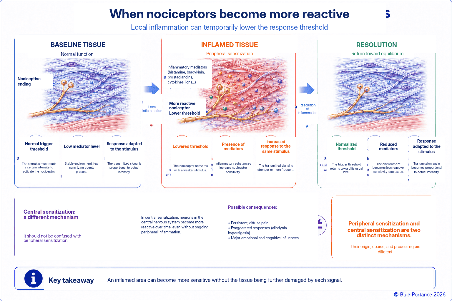
Inflammatory mediators lower the threshold of certain nerve endings. A previously painless stimulus can then become painful, and the sensitive area can widen. This response is biological and potentially reversible. The inflammatory process gradually resolves and the local chemical environment normalizes. The activation threshold of the endings involved can then move back toward its previous level.
An animal model, not a direct transposition
The duration and reversibility of these changes, however, vary with the tissue and with the intensity and persistence of the inflammatory process, and they do not follow a fixed timeline. In a rat model of inflamed thoracolumbar fascia, prolonged experimental inflammation was accompanied by lasting sensitization of dorsal horn neurons. It also increased the density of presumably nociceptive fibers in the tissue (Hoheisel & Mense, 2015), (Hoheisel et al., 2015). These data come from specific experimental conditions in animals; they should therefore not be transposed as is to human fascial inflammation. They nevertheless show that the resolution timeline is not always a simple, rapid return to the initial state.
Primary and secondary hyperalgesia
Two distinct phenomena contribute to this widening — a classic distinction, although one derived largely from experimental models of cutaneous pain (Treede et al., 1992). Primary hyperalgesia refers to increased sensitivity located directly in the inflamed area, where the activation threshold of the nociceptive endings themselves is lowered. Secondary hyperalgesia refers to increased sensitivity in areas outside the initial site. It is attributed mainly to increased excitability and increased central processing of sensory input, particularly at the spinal relays. It is therefore not caused by a pathological state of the tissues in this surrounding area.
Why pain can seem to spread
This distinction explains why a painful area can seem to extend beyond the anatomical boundaries of the tissue that was initially irritated. This extension does not, however, necessarily imply a physical spread of the injury or of the inflammation as such. It also sheds light on why caution is warranted when pain is felt at a distance from an obvious area of mechanical stress. Such pain does not, on its own, prove the existence of fascial continuity “transmitting” pain along an entire anatomical chain. An explanation centered on nervous system sensitization is an alternative explanation to consider, one with solid neurophysiological foundations. It is not possible, however, to state how common it is relative to a mechanical explanation based on altered tissue loading or interfaces.
4. Persistence and central amplification
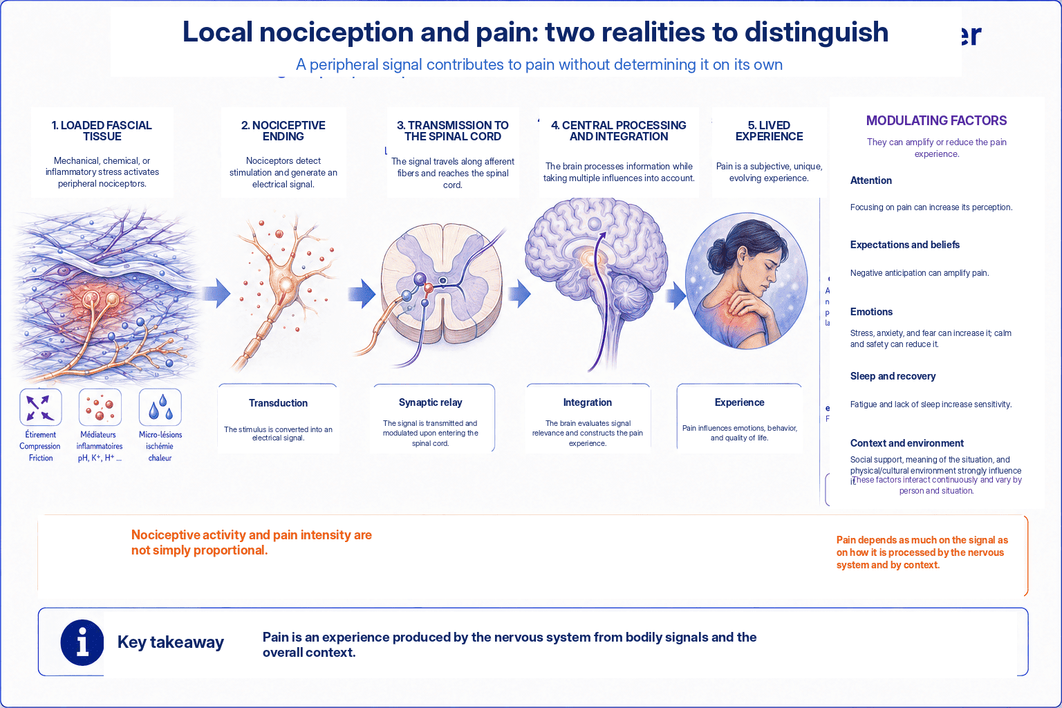
Repeated nociceptive input can alter spinal and brain processing. Pain then becomes less proportional to the local mechanical state. The “Understanding Pain” guide covers these mechanisms in detail. Fascia is thus only one possible peripheral source among others, to be placed within a broader understanding of the mechanisms of persistent pain.
Sufficiently intense, persistent, or repeated nociceptive input can alter excitability and signal processing in spinal and supraspinal circuits. This phenomenon is known as central sensitization (Woolf, 2011). Some of these changes in excitability actually appear quickly in experimental models, without requiring prolonged exposure. The spinal neurons involved in pain processing then become more easily excitable. This change can persist beyond the resolution of the initial peripheral cause.
When the processing system, rather than the tissue, has changed
In this situation, mechanical stimuli that were previously only mildly painful or not painful at all in the region concerned can then trigger a heightened response. This does not reflect further deterioration of the fascia, but a change in the sensitivity of the pain-processing system itself. This general mechanism of central sensitization has not, however, been demonstrated specifically for low, isolated fascial stresses. There is therefore no simple, proportional relationship between identifiable peripheral abnormalities and the intensity of the pain felt. This is one of the most important points for interpreting persistent pain correctly. Beyond a certain stage, searching exclusively for a local mechanical explanation strictly proportional to pain intensity can become a dead end. This holds true even for the most attentive and experienced examiner.
Central sensitization and nociplastic pain: two distinct concepts
This central sensitization should not, however, be confused with so-called nociplastic pain, a more recent and more specific term. Central sensitization refers to a specific neurophysiological mechanism — increased excitability of neurons in the central nervous system. Nociplastic pain is a broader clinical descriptor. It is used to describe pain that arises from altered nociceptive processing. In that case, no clear evidence of tissue damage activating peripheral nociceptors fully explains it. Nor is it fully explained by any lesion or disease of the somatosensory system (Kosek et al., 2016).
A component that can be added without redefining the tissue
Central sensitization can be present in pain whose mechanism is otherwise well identified, without being sufficient on its own to classify that pain as nociplastic. Likewise, chronicity alone does not justify concluding that a mechanism is nociplastic. Pain can remain chronic while still having a clearly identifiable nociceptive or neuropathic cause. Applied to fascia, this distinction means that pain initially associated with identifiable fascial stress can, over time, include a superimposed nociplastic component. The fascia itself does not, strictly speaking, become “nociplastic.” This term describes a pain-processing mechanism, not a property of a peripheral tissue.
5. Why diagnosis remains difficult
The notion of “myofascial pain,” widely used in clinical practice, generally refers to regional pain. This pain is characterized by tender areas located in the muscle or the surrounding fascia. The clinical term “myofascial” should not, however, be interpreted as proof. The anatomical fascia is not necessarily the structure generating the pain. The term refers to a regional clinical syndrome, not a precise tissue diagnosis.
This clinical entity remains debated in the scientific literature. The debate concerns, in particular, its precise diagnostic criteria and the reproducibility of its identification between examiners. Its specificity relative to other forms of regional musculoskeletal pain is also in question.
Systematic reviews find marked heterogeneity in the criteria actually used from one study to the next (Li et al., 2020). Certain signs, however, are an exception, particularly reproduced pain and referred pain. They can reach acceptable interrater reliability after specific training (Bron et al., 2007). The problem therefore lies mainly in the absence of a consensus reference test and in the variability of protocols. It does not call the clinical entity as a whole into question.
A clinical entity without a single biological marker
Myofascial pain lacks specific markers. Perceived stiffness does not prove fibrosis, just as an altered image does not prove causation. Finally, improvement after mobilization does not prove that fascia was the sole source.
Why differential diagnosis remains limited
This difficulty stems from several cumulative reasons. First, current medical imaging cannot directly visualize the functional state of fascial nerve endings or their level of activation. It can, however, detect structural differences at the group level. For example, some people with chronic low back pain show a greater measured thoracolumbar fascia thickness than matched controls (Langevin et al., 2009).
Such a statistical association, observed at the group level, does not, however, allow a conclusion to be drawn. In a specific individual, a given thickness may be the cause, the consequence, or simply an independent correlate of the pain. A group-level abnormality is not an individual diagnostic test.
Three methodological limitations to be aware of
Second, palpation is often used in clinical practice to locate a tender area. It nevertheless loads several superimposed tissues at once: skin, fascia, muscle, periosteum. It therefore cannot isolate the specific contribution of each one.
Finally, symptomatic improvement after a manual or therapeutic intervention does not, on its own, demonstrate that the targeted tissue was the cause of the symptom. It may result from several mechanisms that are not specific to the targeted tissue: an overall change in movement, reduced apprehension, sensory modulation, the person’s expectations. It may also result from the natural course of the symptoms, and this does not by itself validate the initial mechanical hypothesis. This methodological caution does not invalidate the clinical value of a fascia-focused approach. It simply invites us not to turn a plausible hypothesis into diagnostic certainty on the sole basis of a favorable response to treatment.
Key Takeaways
- An innervated fascia can contribute to nociception.
- Pain can come from the tissue or from its interfaces.
- Inflammation and sensitization alter the response threshold.
- Chronic pain cannot be attributed to fascia on the basis of a single clue.
Frequently Asked Questions
Can pain without a visible lesion be fascial?
Can palpation identify a painful fascia?
Can pain initially associated with fascia later include a nociplastic component?
Does improvement after fascial treatment prove that fascia was the cause?
Scientific References
- Suarez-Rodriguez V, Fede C, Pirri C, et al. Fascial innervation: a systematic review. Int J Mol Sci. 2022;23:5674.
- Schilder A, Hoheisel U, Magerl W, et al. Sensory findings after stimulation of the thoracolumbar fascia. Pain. 2014;155:222–231.
- Woolf CJ. Central sensitization: implications for the diagnosis and treatment of pain. Pain. 2011;152(3 Suppl):S2–S15.
- Hoheisel U, Mense S. Inflammation of the thoracolumbar fascia excites and sensitizes rat dorsal horn neurons. Eur J Pain. 2015;19(3):419–428.
- Hoheisel U, Rosner J, Mense S. Innervation changes induced by inflammation of the rat thoracolumbar fascia. Neuroscience. 2015;300:351–359.
- Treede RD, Meyer RA, Raja SN, Campbell JN. Peripheral and central mechanisms of cutaneous hyperalgesia. Prog Neurobiol. 1992;38(4):397–421.
- Kosek E, Cohen M, Baron R, et al. Do we need a third mechanistic descriptor for chronic pain states? Pain. 2016;157(7):1382–1386.
- Langevin HM, Stevens-Tuttle D, Fox JR, et al. Ultrasound evidence of altered lumbar connective tissue structure in human subjects with chronic low back pain. BMC Musculoskelet Disord. 2009;10:151.
- Bron C, Franssen J, Wensing M, Oostendorp RAB. Interrater reliability of palpation of myofascial trigger points in three shoulder muscles. J Man Manip Ther. 2007;15(4):203–215.
- Li L, Stoop R, Clijsen R, et al. Criteria used for the diagnosis of myofascial trigger points in clinical trials on physical therapy: updated systematic review. Clin J Pain. 2020;36(12):955–967.
← Previous chapter: Is Fascia a Neurosensory Interface?
Back to the Fascia guide
Next chapter: How Can Fascia Become Altered? Inflammation, Densification, and Fibrosis →
Note: this content explains general mechanisms. It does not constitute a medical diagnosis or a therapeutic recommendation.
© Gil Ayache. The original concepts, biomechanical models, diagrams, terminology, graphic representations, foundational figures, original texts, and principles presented on this page are works protected by copyright. They are made available to Blue Portance under an intellectual property license agreement, without transfer of economic rights or of authorship.
