


This article is part of the Expert Guide “Fascia.”
Chapter 7 — Blue Portance Knowledge BaseIs Fascia a Neurosensory Interface?
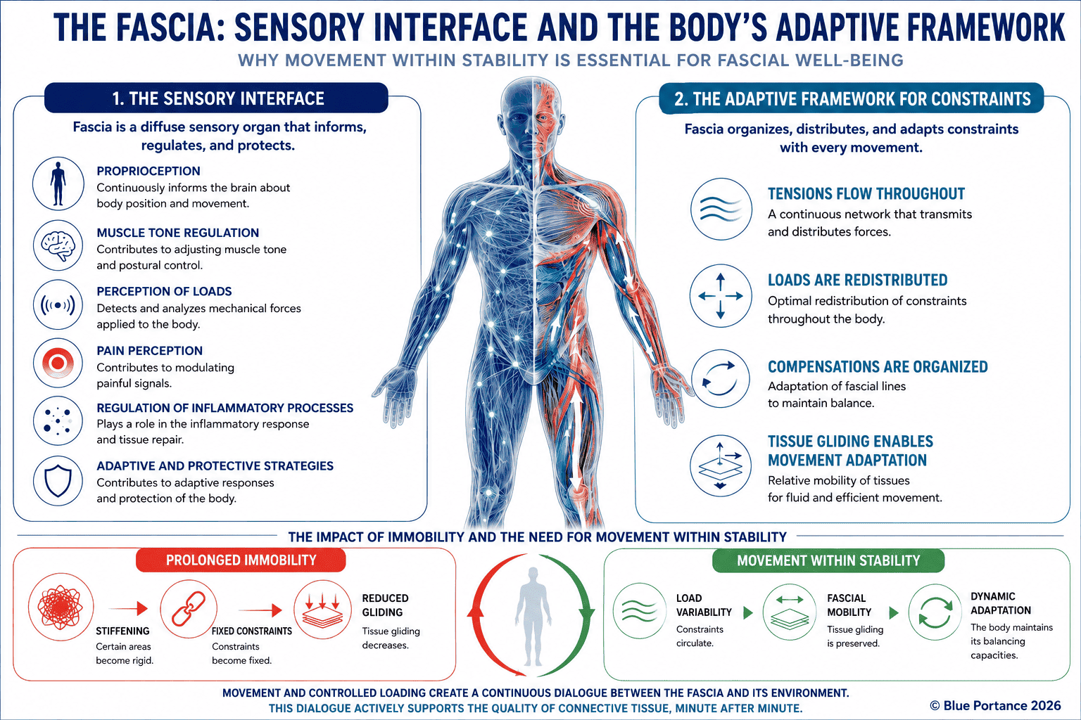
Summary — Fascia contains sensory and autonomic nerve fibers at varying densities. Free nerve endings can respond to mechanical or chemical stimuli. Other, more specialized receptors contribute to information about position and movement. Fascia thus forms an interface where the mechanical and biological state of the tissue becomes neural information. The central nervous system transmits, modulates, and then integrates this information.
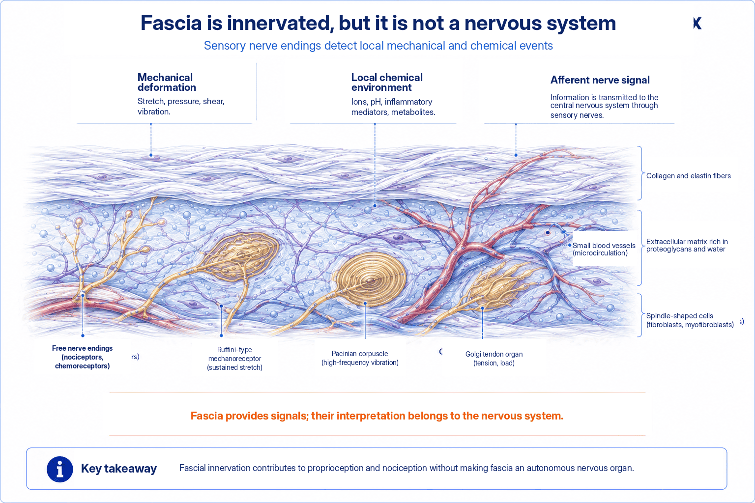
1. Heterogeneous Innervation
Histological studies have identified nerve fibers in several fasciae, notably the thoracolumbar fascia. The superficial layer and areas close to blood vessels or attachments often show denser innervation than other areas (Suarez-Rodriguez et al., 2022).
This innervation combines several types of nerve endings. A landmark histological study of human specimens thus reported two types of encapsulated corpuscles alongside free nerve endings. These are Ruffini corpuscles and Vater-Pacini corpuscles (Yahia et al., 1992). A later study explored the same question quantitatively in rats, and only on a preliminary basis in human specimens. It did not find these encapsulated corpuscles in its own human sample (Tesarz et al., 2011).
This divergence thus shows how much the species studied, the sample size, and the quantification protocol can influence what is actually reported from one study to the next. Encapsulated corpuscles, sensitive to sustained stretch or to rapid vibration depending on their type, thus coexist with a dense network of free, unencapsulated nerve endings. This coexistence varies across studies and across the fasciae examined, and these free nerve endings remain harder to classify functionally.
A Distribution That Varies with Depth
The density and relative proportion of these different types vary not only from one fascia to another but also with depth within the same fascia. The most superficial layers and the areas close to tendon attachments or blood vessels are thus generally the most richly innervated. This heterogeneity makes any generalization such as “fascia is innervated” hazardous. Indeed, one needs to specify which fascia is being discussed, at what depth, and using which study method.
A Regional Contrast Documented in Animals
In mice, for example, detailed mapping demonstrated that the thoracolumbar fascia is roughly three times more innervated than the gluteal fascia (Fede et al., 2021). This contrast, visible both in surface area and in branching density, clearly illustrates this regional heterogeneity. Nevertheless, these precise values, obtained in animals, should not be transposed as such to humans. The gradual recognition of this sensory richness has contributed greatly to changing how fascia is represented. Long reduced to a mere supporting tissue, fascial tissues are increasingly recognized as contributors to body perception (Lesondak, 2019).
2. Mechanoreception and Proprioception
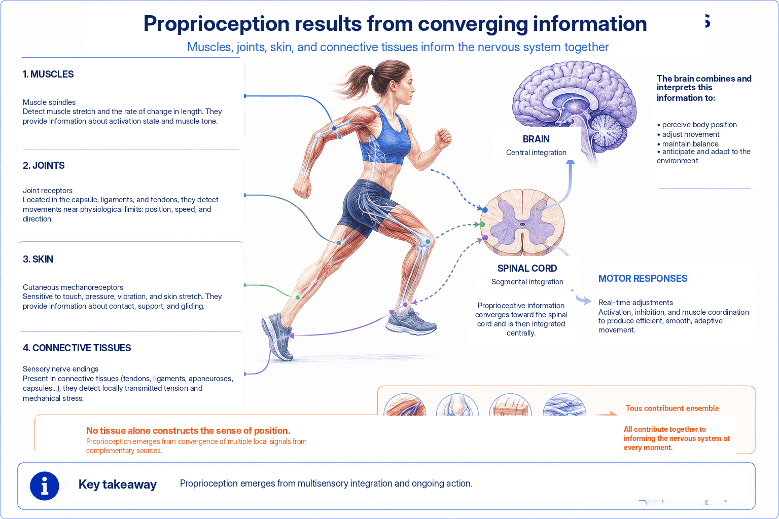
Tissue deformation contributes to the somatosensory information used to adjust posture and movement. However, in a real-world task, it is difficult to isolate the contribution of fascia from that of muscles, joints, and skin.
Proprioception — the perception of the body’s position and movement — classically relies on the convergence of information. This information comes from muscle spindles, joint receptors, the skin, and, to a degree still under debate, fascial tissues themselves. Reference models in sensory physiology emphasize precisely this population-based, convergent nature of proprioceptive information. No single receptor encodes a position or a movement. Rather, it is the integration of discharges from several classes of receptors activated simultaneously that does so (Proske & Gandevia, 2012).
A Contribution Hard to Isolate from the Rest of the Body
A receptor located in fascia near a joint remains mechanically coupled to neighboring structures. Direct stretching of the fascial tissue and the movements of these structures can thus both stimulate it. Outside highly controlled experimental conditions, this makes it very difficult to attribute a given piece of proprioceptive information to a single tissue source. This methodological difficulty explains the caution in the scientific literature regarding the exact quantitative contribution of fascia to overall proprioception. Its qualitative contribution, however, is not called into question.
When sitting, this convergence of information is in particularly high demand. Posture must indeed be adjusted continuously based on signals from the pelvis, the lower limbs, the trunk, and the contact surface with the seat itself. Some of these signals pass through the connective tissues surrounding the joint and muscle structures involved. Fascia thus becomes one contributor among others to the overall perception of seated posture. However, given current knowledge, it is not possible to isolate precisely its share relative to the other somatosensory tissues engaged in the same task. This methodological limitation does not diminish the practical relevance of the observation. A seat that modulates the deformation of the surrounding connective tissues thus, by definition, modulates part of the somatosensory information available for postural adjustment. The exact extent of this contribution, however, remains to be clarified by future research.
3. Free Nerve Endings and Nociception
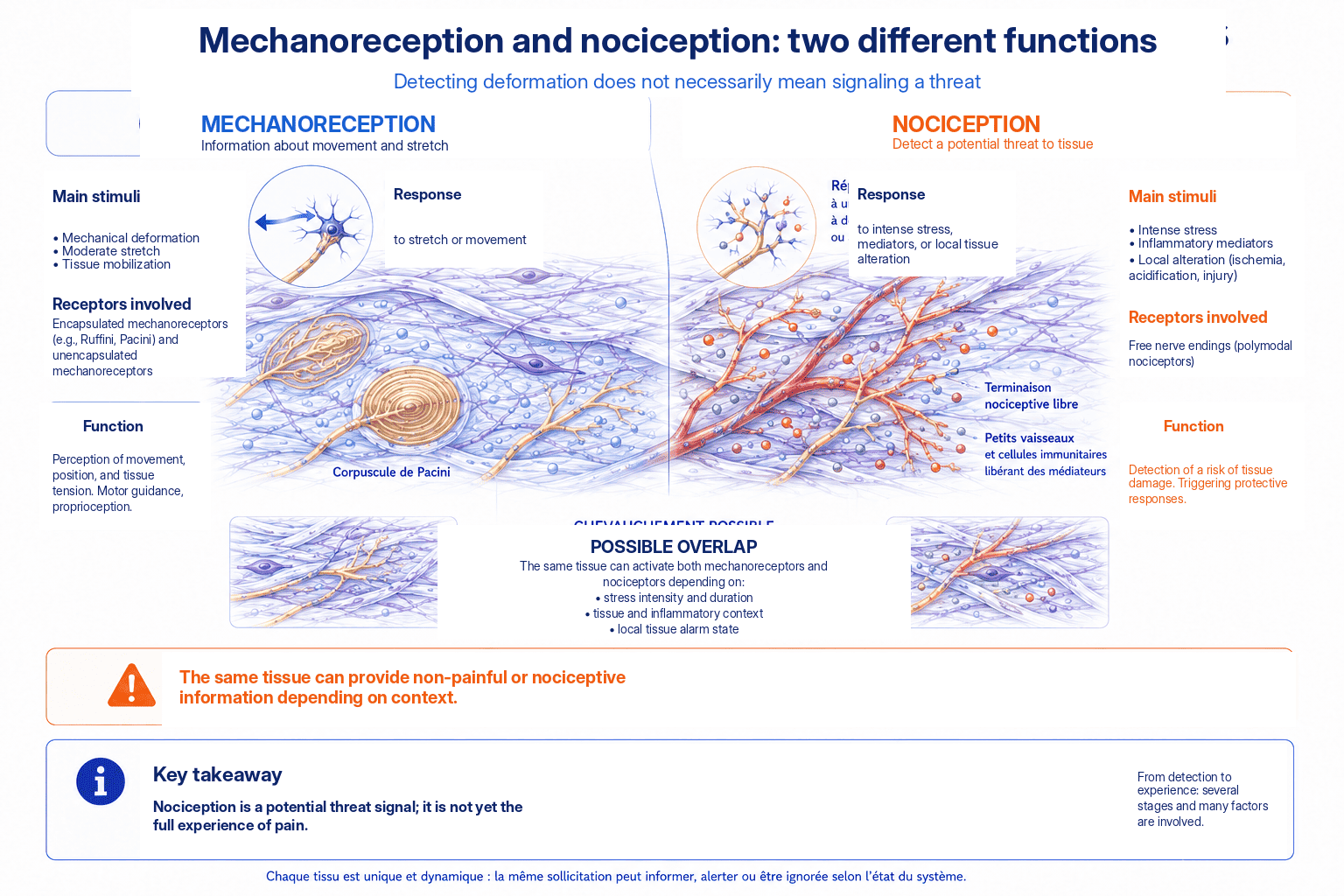
Free nerve endings can respond to potentially harmful stimuli. An experimental injection into the thoracolumbar fascia can produce widespread, long-lasting pain. This thus shows its capacity to contribute to nociception (Schilder et al., 2014).
This experimental protocol made it possible to compare the sensitivity of different tissue layers by injecting hypertonic saline at different depths. The results showed that injection into the thoracolumbar fascia could produce more intense and longer-lasting pain than the same injection into the underlying muscle. This therefore suggests a particularly marked nociceptive sensitivity of this connective tissue (Schilder et al., 2014). However, these specific experimental conditions and this particular fascia limit the scope of this finding. It should therefore not be generalized without caution to all the fasciae of the body, nor taken as proof that all diffuse pain necessarily has a fascial origin.
Peripheral and Central Sensitization
The chemical mechanisms involved in this sensitization deserve clarification. Local irritation or inflammation is accompanied by the release of several mediators, including prostaglandins, bradykinin, and various cytokines. These mediators can thus lower the activation threshold of surrounding nociceptive nerve endings (Pethő & Reeh, 2012). This phenomenon, called peripheral sensitization, explains why an inflamed area temporarily becomes more sensitive to stimuli. Under normal circumstances, these stimuli would indeed not be perceived as painful. This is not a property exclusive to fascial tissue. The same mechanism is indeed observed in most innervated tissues subjected to local inflammation.
This peripheral sensitization can itself extend into the central nervous system. Repeatedly stimulated spinal and supraspinal relays then become more reactive to stimuli they would not previously have perceived as painful. This distinct phenomenon, called central sensitization (Woolf, 2011) and discussed in the next chapter, helps explain some persistent pain that is disproportionate to the tissue injury originally involved.
4. Mechanical Interactions with Nerves
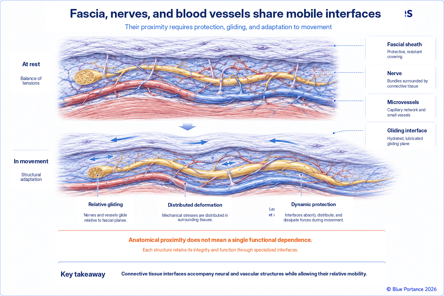
Fascia also forms the external environment of nerves. A loss of gliding can increase tension or shear without fascia itself being the sole generator of the signal. The mechanical interface must be distinguished from the sensory interface.
Chapter 4 showed that a peripheral nerve must be able to glide within its connective tissue environment to accompany movement. It thus avoids being exposed to excessive stretch or compression (Topp & Boyd, 2006). This gliding may decrease, for example if the surrounding connective tissue becomes locally more adherent or less mobile. The nerve may then be subjected to increased mechanical tension with each movement. This in turn can activate its own sensory fibers. The peripheral nerve indeed has its own intrinsic sensory and sympathetic innervation, housed in its connective tissue sheaths (epineurium, perineurium, endoneurium). These fibers, known as the nervi nervorum, can thus become a source of nociception in their own right when the nerve itself is mechanically irritated. This remains independent of any damage to the axons it carries (Bove & Light, 1997).
Two Roles Not to Be Confused
This situation illustrates why it is important to distinguish two different roles that fascia can play. It can be a source of sensory signals through its own nerve endings, or simply a mechanical environment whose state alters the loading of a neighboring nerve. Confusing these two roles leads to oversimplified explanations of a mechanism that in reality operates on two levels. This distinction has practical implications. Discomfort felt along the course of a nerve may thus arise from direct activation of the nerve’s own sensory fibers. It may also result from excessive mechanical loading transmitted to it by a less mobile connective tissue environment, or from a combination of both. This therefore explains why the exact origin of a peripheral nerve sensation often remains difficult to determine without a thorough examination and an appropriate clinical evaluation.
5. From Peripheral Signal to Perception
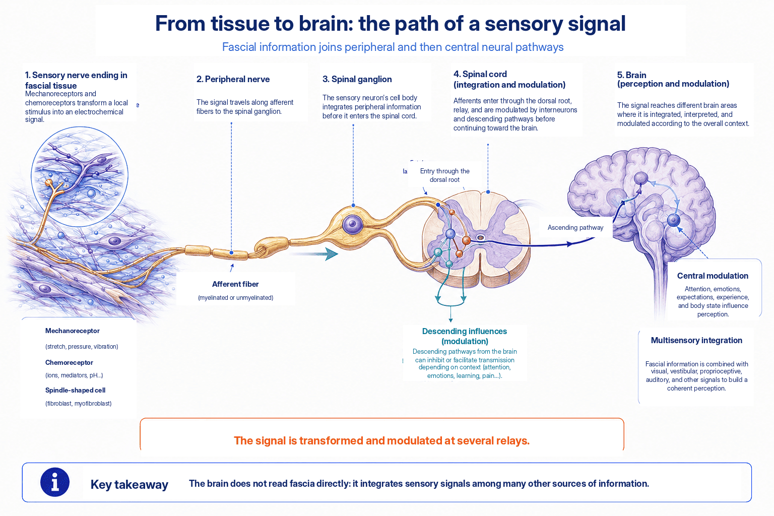
Tissue does not “feel” on its own. Nerve pathways transmit these signals, the spinal cord modulates them, and the brain then integrates them with context, attention, and experience. Fascial input is therefore not automatically equivalent to pain.
Between the activation of a peripheral nerve ending and the conscious perception of a sensation, several stages of processing and modulation come into play. In the spinal cord, other sensory information present at the same time can amplify or dampen the signal. Descending pathways from the brainstem are also involved. Depending on the context, they can facilitate or, conversely, inhibit the transmission of the nociceptive message to higher centers (Ossipov et al., 2010). In the brain, final integration draws on areas associated with attention, emotional state, and the person’s past experience. All of these areas thus influence how the same peripheral signal is ultimately interpreted.
This multi-level pathway explains why the same mechanical loading of a fascia can produce very different responses depending on the context. It may thus produce no conscious sensation at all, a simple perception of tension, or outright pain. None of these responses, however, directly reflects on its own the state of the peripheral tissue. It is this integrative dimension, developed in more detail in the next chapter, that makes it necessary to distinguish two notions. On the one hand, a peripheral nociceptive signal, biologically real and measurable. On the other hand, a conscious experience of pain, which always results from processing at several levels of the nervous system.
Key Takeaways
- Fascial innervation varies considerably.
- Fascia contributes to mechanical and nociceptive information.
- The nervous system transforms these signals into perception.
- Fascia should not be presented as an autonomous peripheral brain.
Frequently Asked Questions
Can fascia hurt?
Is fascia more innervated than muscle?
Does diffuse pain prove a fascial origin?
Does fascia really contribute to proprioception?
Scientific References
- Suarez-Rodriguez V, Fede C, Pirri C, et al. Fascial innervation: a systematic review. Int J Mol Sci. 2022;23:5674.
- Yahia L, Rhalmi S, Newman N, Isler M. Sensory innervation of human thoracolumbar fascia. Acta Orthop Scand. 1992;63:195–197.
- Tesarz J, Hoheisel U, Wiedenhöfer B, Mense S. Sensory innervation of the thoracolumbar fascia in rats and humans. Neuroscience. 2011;194:302–308.
- Schilder A, Hoheisel U, Magerl W, et al. Sensory findings after stimulation of the thoracolumbar fascia. Pain. 2014;155:222–231.
- Lesondak D. Le Fascia. [Fascia.] Éditions Ressources Primordiales; 2019.
- Fede C, Petrelli L, Guidolin D, et al. Evidence of a new hidden neural network into deep fasciae. Sci Rep. 2021;11:12623.
- Proske U, Gandevia SC. The proprioceptive senses: their roles in signaling body shape, body position and movement, and muscle force. Physiol Rev. 2012;92(4):1651–1697.
- Pethő G, Reeh PW. Sensory and signaling mechanisms of bradykinin, eicosanoids, platelet-activating factor, and nitric oxide in peripheral nociceptors. Physiol Rev. 2012;92(4):1699–1775.
- Woolf CJ. Central sensitization: implications for the diagnosis and treatment of pain. Pain. 2011;152(3 Suppl):S2–S15.
- Topp KS, Boyd BS. Structure and biomechanics of peripheral nerves: nerve responses to physical stresses and implications for physical therapist practice. Phys Ther. 2006;86(1):92–109.
- Bove GM, Light AR. The nervi nervorum: missing link for neuropathic pain? Pain Forum. 1997;6(3):181–190.
- Ossipov MH, Dussor GO, Porreca F. Central modulation of pain. J Clin Invest. 2010;120(11):3779–3787.
← Previous chapter: Mechanotransduction: How Does Stress Change Fascial Tissue?
Back to the Fascia guide
Next chapter: How Can Fascia Contribute to Pain? →
Note: this content explains general mechanisms. It does not constitute a medical diagnosis or a therapeutic recommendation.
© Gil Ayache. The original concepts, biomechanical models, diagrams, terminology, graphic representations, foundational figures, original texts, and principles presented on this page are works protected by copyright. They are made available to Blue Portance under an intellectual property license agreement, without transfer of economic rights or of authorship.
