


This article is part of the Expert Guide “Fascia.”
Chapter 6 — Blue Portance Knowledge BaseMechanotransduction: How Does Stress Change Fascial Tissue?
Summary — Cells are not isolated from their mechanical environment. A deformation of the matrix can be transmitted through integrins to the cytoskeleton, then alter signaling pathways and gene expression. Fibroblasts then adjust the production, organization, and breakdown of the matrix. This passage from mechanics to biology is what is called mechanotransduction. It is a central mechanism for understanding how a tissue adapts—or maladapts—to its mechanical environment over time.
1. From the Matrix to the Cell
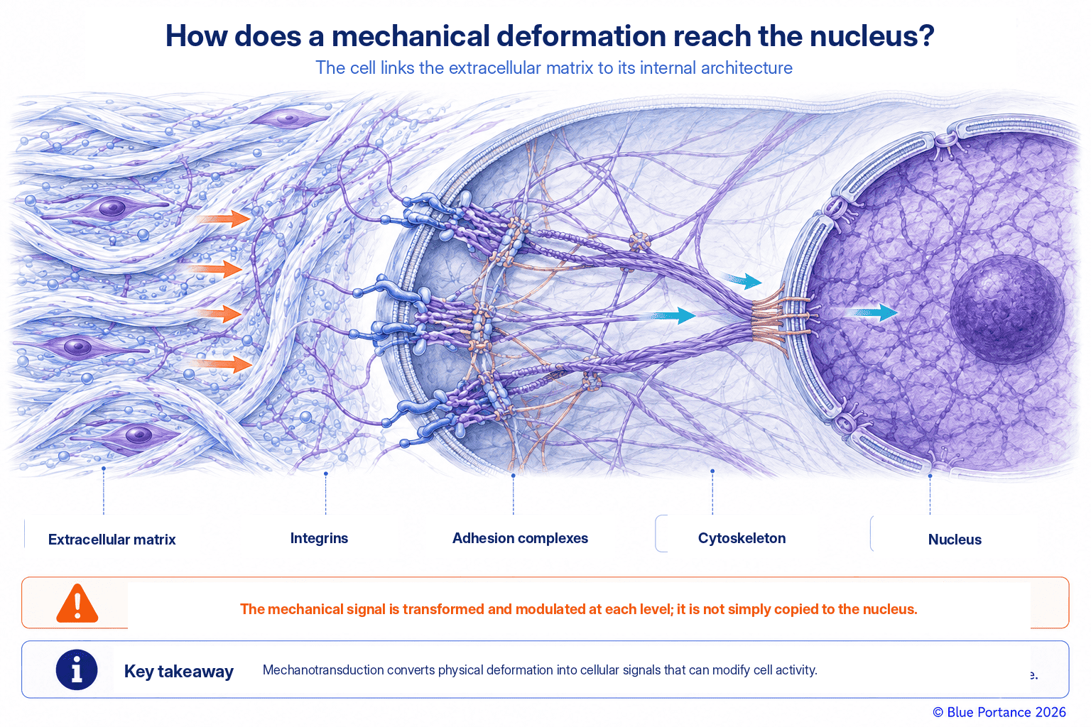
Integrins link the extracellular matrix to the cytoskeleton. When the matrix deforms, adhesion complexes, mechanosensitive channels, and cytoskeletal tension respond. Together, they can alter cellular activity (Chiquet et al., 2009).
This chain of transmission does not follow a single, sequential pathway. Rather, it relies on several relays that operate in parallel and interact with one another. Integrins thus cluster at focal adhesion sites. There, they associate with a set of proteins, including kinases such as FAK. These proteins physically connect the cell membrane to the actin network of the cytoskeleton while simultaneously transmitting biochemical signals (Chiquet et al., 2009). A deformation of the matrix therefore changes the tension exerted on this network. This can in turn open stretch-sensitive ion channels and locally alter the concentrations of certain ions, notably calcium.
In parallel, the reorganization of actin itself changes the internal tension of the cell. This can in turn activate transcription factors that are sensitive to mechanical forces.
All the Way to the Nucleus: The LINC Complex
Some of these signals also travel to the nucleus by a distinct route. The cytoskeleton remains physically connected to the nuclear envelope by a dedicated protein complex, the LINC complex (nesprins and SUN proteins). This complex thus allows a mechanical deformation to directly influence chromatin organization. Ultimately, it alters the expression of certain genes (Crisp et al., 2006). These pathways are numerous: focal adhesions, cytoskeletal tension, mechanosensitive channels, mechanosensitive transcription factors, and nucleus–cytoskeleton coupling. They converge rather than follow one another in a strict order. This explains the robustness and speed of cellular mechanotransduction (Ingber, 2003).
A Convergent Chain That Sets the Stage for the Neurosensory Interface
This physical continuity, from the matrix all the way to the nucleus, explains why we speak of mechanotransduction. It is not simply a chemical response to an external signal. These mechanisms, described in many cell types, thus show that connective tissue cells can detect the mechanical properties of their environment. They can also modify their activity accordingly. They therefore help us understand fascia as a biologically responsive tissue, without attributing to it an autonomous capacity for information processing. This general reading of fascia as an active tissue is also the one synthesized by Lesondak (Lesondak, 2019). This reinterpretation of fascia as a biologically responsive tissue, rather than as mere anatomical packaging, thus sets the stage for the next chapter. That chapter is devoted to its role as a neurosensory interface.
2. The Fibroblast as Organizer of the Matrix
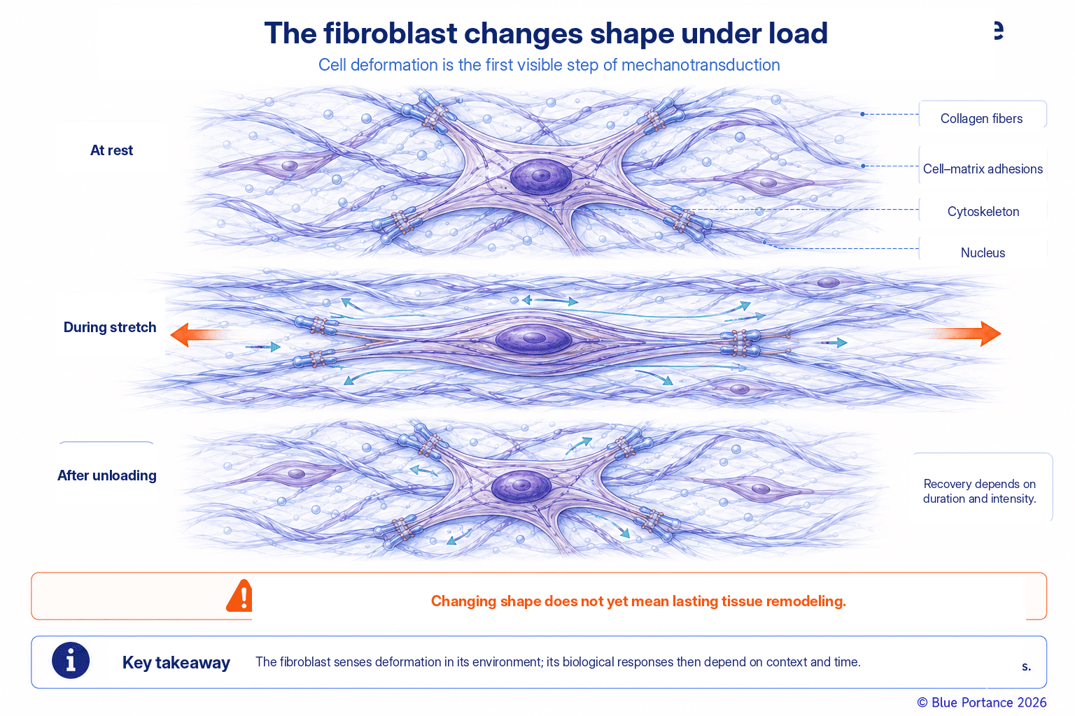
The fibroblast synthesizes collagen, proteoglycans, and remodeling enzymes. In mice, stretching subcutaneous tissue can change the shape of fibroblasts within minutes. This morphological response is far faster than would be expected from the synthesis of new proteins (Langevin et al., 2005).
Direct observations were made ex vivo and in vivo on mouse subcutaneous tissue stretched for periods ranging from ten minutes to two hours. They showed that the fibroblast cytoskeleton rapidly reorganizes in response to stretch. This reorganization is accompanied by a measurable increase in cell perimeter and cell area (Langevin et al., 2005). This study documents a murine model and a cellular morphological response. However, it does not directly measure new collagen production or lasting matrix remodeling. It therefore demonstrates neither human fascial adaptation nor the clinical effect of mobilization.
This finding nevertheless suggests that fibroblasts do not merely build the matrix passively: they actively respond to its mechanical state. This rapid cytoskeletal response thus precedes, in time, the slower adjustment of their synthetic activity.
A Dual Capacity, Fast and Slow
This dual capacity—a fast change in shape and a slow change in synthesis—allows the fibroblast to act as a continuous organizer of the matrix. It is not merely an occasional producer. In many connective tissues, it thus adjusts both the amount of collagen produced and the type of collagen synthesized. It also regulates the activity of the enzymes that break down old matrix (matrix metalloproteinases), as well as that of their inhibitors. This regulation of matrix gene expression is documented in the review by Chiquet and colleagues (Chiquet et al., 2009).
This balancing act goes on continuously, even in the absence of any disorder or particular event. Mechanical conditions contribute to regulating this balance. Their effect, however, depends on the cell type, the tissue, and the characteristics of the loading—intensity, duration, frequency, and mode. It also depends on the local biochemical context, notably the presence of growth factors or inflammatory signals. The same deformation therefore does not necessarily produce the same response in all fasciae or in all individuals.
Heterogeneity Among Fibroblasts
This variability stems largely from the heterogeneity of fibroblasts themselves. Far from forming a uniform cell population, fibroblasts differ according to their embryonic origin and to the tissue and anatomical region in which they reside. They thus retain lasting functional differences even in culture (Plikus et al., 2021). A fibroblast from superficial fascia, a tendon fibroblast, and a fibroblast from deep fascia therefore do not necessarily share the same mechanical sensitivity. Nor do they share the same rate of matrix turnover or the same threshold for switching to a myofibroblast phenotype. This heterogeneity, still incompletely mapped across the different tissues of the fascial system, therefore calls for caution. A result obtained in one tissue or one species should not be extended to all human fascial tissues.
3. Two Timescales Not to Be Confused
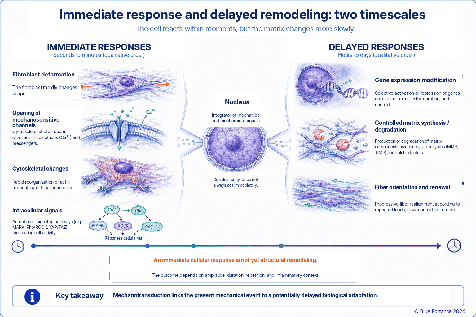
The displacement of a fluid or the gliding of a layer is immediate. A lasting change in fibrous organization requires cellular activity, synthesis, and remodeling that unfold over days, weeks, or longer.
This distinction echoes the one drawn in Chapter 2 between the immediate mechanical response—creep, stress relaxation, hysteresis—and delayed biological adaptation. A connective tissue that is regularly and progressively loaded can thus change over weeks to months. Its collagen content, fiber orientation, and overall stiffness may be modified as a result. This adaptation is particularly well documented in humans in tendons and aponeuroses. There, mechanical loading alters collagen synthesis and the mechanical properties of the tissue (Magnusson et al., 2008). It thus provides a useful model for understanding the remodeling of connective tissues under load.
This model does not, however, justify automatically attributing the same time frames or the same magnitude of response to all components of the fascial system. Their cellular heterogeneity has, moreover, just been highlighted.
A Model That Does Not Apply Uniformly
The effects of reduced loading are not uniform across tissues either. A short period of immobilization can thus markedly reduce muscle mass and strength. However, it does not measurably change tendon collagen turnover over the same period. This illustrates that muscle and dense connective tissue do not respond at the same speed to the same stress (Christensen et al., 2008). Confusing these timescales leads to unrealistic expectations: a mechanical deformation or cellular signaling can be triggered within minutes. A lasting structural transformation of the matrix, however, requires repetition of the stimulus. It also requires sufficient turnover time, on the order of several weeks to several months.
Do Not Confuse the Speed of the Signal with That of Remodeling
It is this consistent repetition of a stimulus that determines observable biological remodeling. This principle thus applies as much to favorable adaptation to progressive training as to maladaptation linked to prolonged sedentary behavior. This timescale is a reason to put changes perceived in the moment into perspective. A feeling of stiffness or suppleness experienced within a few minutes mainly reflects the immediate mechanics described above, not biological remodeling that has already taken place.
4. Adaptation or Maladaptation
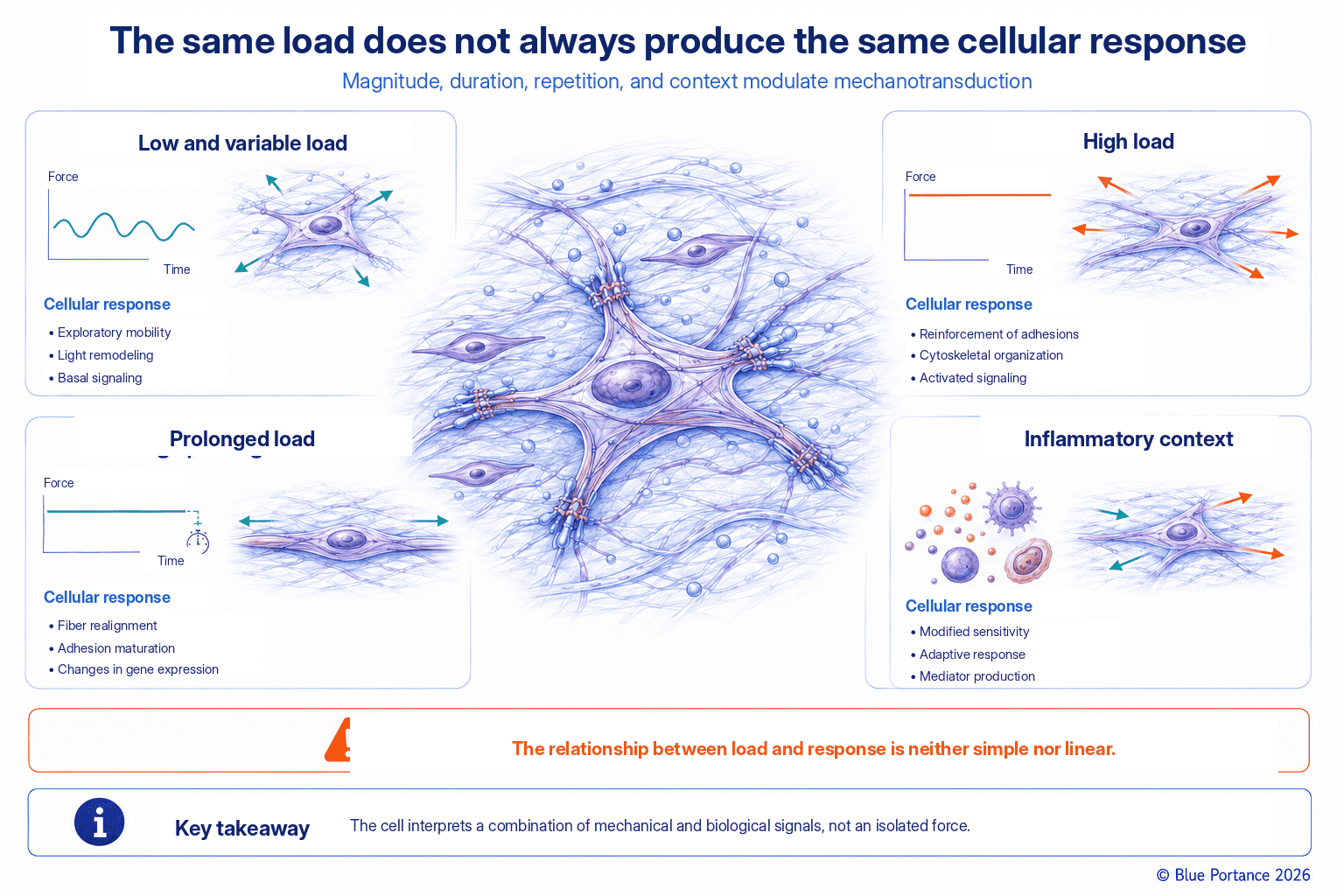
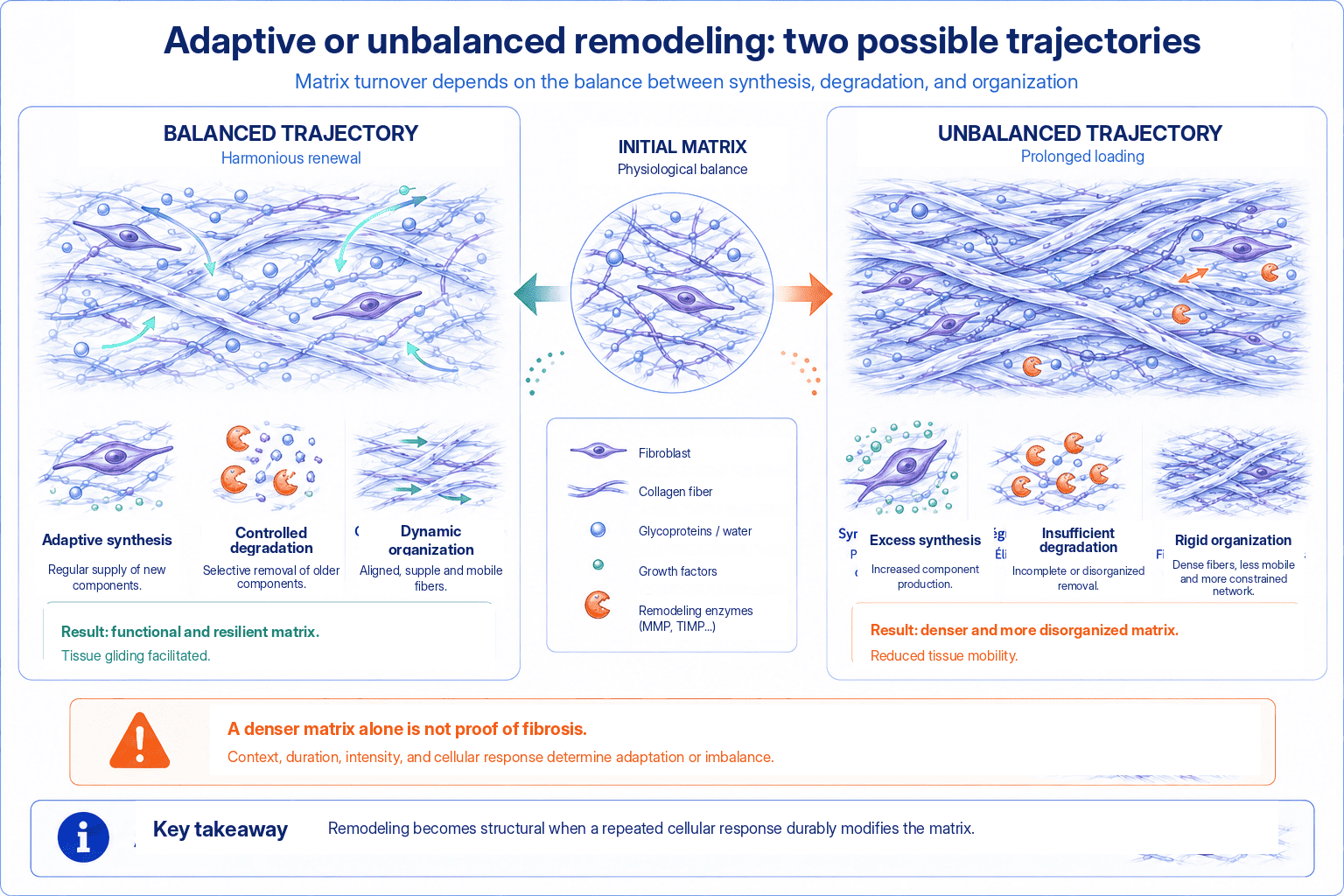
In certain connective tissues, progressive loading can modify the synthesis, organization, and mechanical properties of the matrix. This remodeling then follows the direction of the repeated loads. Excessive or prolonged loading, or loading combined with inflammation, can promote myofibroblasts, collagen deposition, and stiffening. Mechanotransduction is therefore neither good nor bad in itself.
This fork between two opposite outcomes rests on the same basic cellular mechanism. It is one of the most important points to take away from this chapter. The direction this cellular response takes depends on several combined factors. Among them are the intensity of the load relative to the tissue’s current capacities, its frequency and repetition, and the presence or absence of sufficient recovery periods. The local biological context also matters—notably whether or not there is associated inflammation.
The same cell can thus respond adaptively to a progressive, moderate load, helping to maintain a matrix organized along the usual lines of force. This alone, however, does not guarantee the absence of pain or the restoration of function. It can also respond maladaptively to a load that is too intense, too sustained, or superimposed on an inflammatory context.
What Determines Which Way It Tips
In the latter case, matrix tension interacts with biochemical signals, foremost among them TGF-β. This coupling thus promotes the differentiation of fibroblasts into contractile myofibroblasts. This joint mechanoregulation of tension and growth factors is well documented. The literature on wound healing and fibrosis confirms it (Tomasek et al., 2002). It is this possible tipping, in one direction or the other, that directly links cellular mechanotransduction to the fibrotic processes discussed in Chapter 9.
The Myofibroblast, a Central Player in This Ambivalence
The myofibroblast illustrates this ambivalence well. Normally absent or rare in many uninjured adult tissues, it appears transiently during repair, where its contractile capacity helps close a wound. Certain related contractile populations also exist in specialized locations. If, however, it persists in the tissue for an abnormally long time, this same contractile capacity changes role. Combined with excessive deposition and abnormal remodeling of the matrix, it can then contribute to pathological stiffening (Tomasek et al., 2002).
5. A Mechanical and Biological Loop
The matrix influences the cell, which in turn modifies the matrix. Increased stiffness can reinforce certain cellular tensions and sustain remodeling. This loop will be revisited in the chapter on fibrosis.
This loop has an important property: it can become self-sustaining. Increased matrix stiffness can change the forces generated at focal adhesions. In certain contexts, it can thus promote the persistence of a myofibroblast phenotype. Cells then detect this stiffness notably via the mechanosensitive transcription factors YAP and TAZ. The nuclear localization of these factors increases on a stiff substrate (Dupont et al., 2011).
This coupling between stiffening and cellular activation is well documented in specific fibrotic tissue models. There, matrix accumulation in turn sustains stiffness and myofibroblast differentiation. This mechanism partly explains the following phenomenon: certain forms of tissue remodeling tend to continue beyond the initial stimulus that triggered them (Huang et al., 2012). Conversely, mechanical loading that is compatible with the tissue’s capacities can help maintain its turnover. In certain tissues, it can also promote the adaptation of its mechanical properties.
A Less Well-Characterized Adaptive Trajectory
This is not, however, simply the reverse of the fibrotic loop. This trajectory is considerably less precisely characterized in the literature. The cellular pathways, time frames, and effects also depend heavily on the tissue studied. This asymmetry between a well-described pathological loop and an adaptive loop that is harder to pin down is itself an important point. It is a reminder that biology does not necessarily operate along two perfectly symmetrical trajectories. Mechanotransduction remains central to understanding fascial tissues precisely because its outcome is never a foregone conclusion.
Key Takeaways
- Mechanotransduction converts a mechanical signal into a cellular response.
- Fibroblasts modify the matrix according to context.
- Immediate mechanical effects and delayed biological remodeling are distinct.
- The same pathway can support adaptation or contribute to fibrosis.
Frequently Asked Questions
Does movement immediately remodel fascia?
Do fibroblasts sense stiffness?
Does mechanotransduction prove that a treatment works?
Does a heavier load produce greater adaptation?
Scientific References
Cited References (1/2)
- Chiquet M, Gelman L, Lutz R, Maier S. From mechanotransduction to extracellular matrix gene expression. Biochim Biophys Acta. 2009;1793:911–920.
- Christensen B, Dyrberg E, Aagaard P, Kjaer M, Langberg H. Short-term immobilization and recovery affect skeletal muscle but not collagen tissue turnover in humans. J Appl Physiol. 2008;105(6):1845–1851.
- Crisp M, Liu Q, Roux K, et al. Coupling of the nucleus and cytoplasm: role of the LINC complex. J Cell Biol. 2006;172(1):41–53.
- Dupont S, Morsut L, Aragona M, et al. Role of YAP/TAZ in mechanotransduction. Nature. 2011;474(7350):179–183.
- Huang X, Yang N, Fiore VF, et al. Matrix stiffness-induced myofibroblast differentiation is mediated by intrinsic mechanotransduction. Am J Respir Cell Mol Biol. 2012;47(3):340–348.
Cited References (2/2)
- Ingber DE. Tensegrity I. Cell structure and hierarchical systems biology. J Cell Sci. 2003;116:1157–1173.
- Langevin HM, Bouffard NA, Badger GJ, Iatridis JC, Howe AK. Dynamic fibroblast cytoskeletal response to subcutaneous tissue stretch ex vivo and in vivo. Am J Physiol Cell Physiol. 2005;288(3):C747–C756.
- Magnusson SP, Narici MV, Maganaris CN, Kjaer M. Human tendon behaviour and adaptation, in vivo. J Physiol. 2008;586(1):71–81.
- Plikus MV, Wang X, Sinha S, et al. Fibroblasts: origins, definitions, and functions in health and disease. Cell. 2021;184(15):3852–3872.
- Tomasek JJ, Gabbiani G, Hinz B, Chaponnier C, Brown RA. Myofibroblasts and mechano-regulation of connective tissue remodelling. Nat Rev Mol Cell Biol. 2002;3:349–363.
- Lesondak D. Le Fascia. [Fascia.] Éditions Ressources Primordiales; 2019.
← Previous chapter: Why Does Fascia Need Micro-Movements?
Back to the Fascia guide
Next chapter: Is Fascia a Neurosensory Interface? →
Note: this content explains general mechanisms. It does not constitute a medical diagnosis or a therapeutic recommendation.
© Gil Ayache. The original concepts, biomechanical models, diagrams, terminology, graphic representations, foundational figures, original texts, and principles presented on this page are works protected by copyright. They are made available to Blue Portance under an intellectual property license agreement, without transfer of economic rights or of authorship.
