


This article is part of the Expert Guide “Fascia.”
Chapter 1 — Blue Portance Knowledge BaseWhat Is Fascia and How Is It Organized?
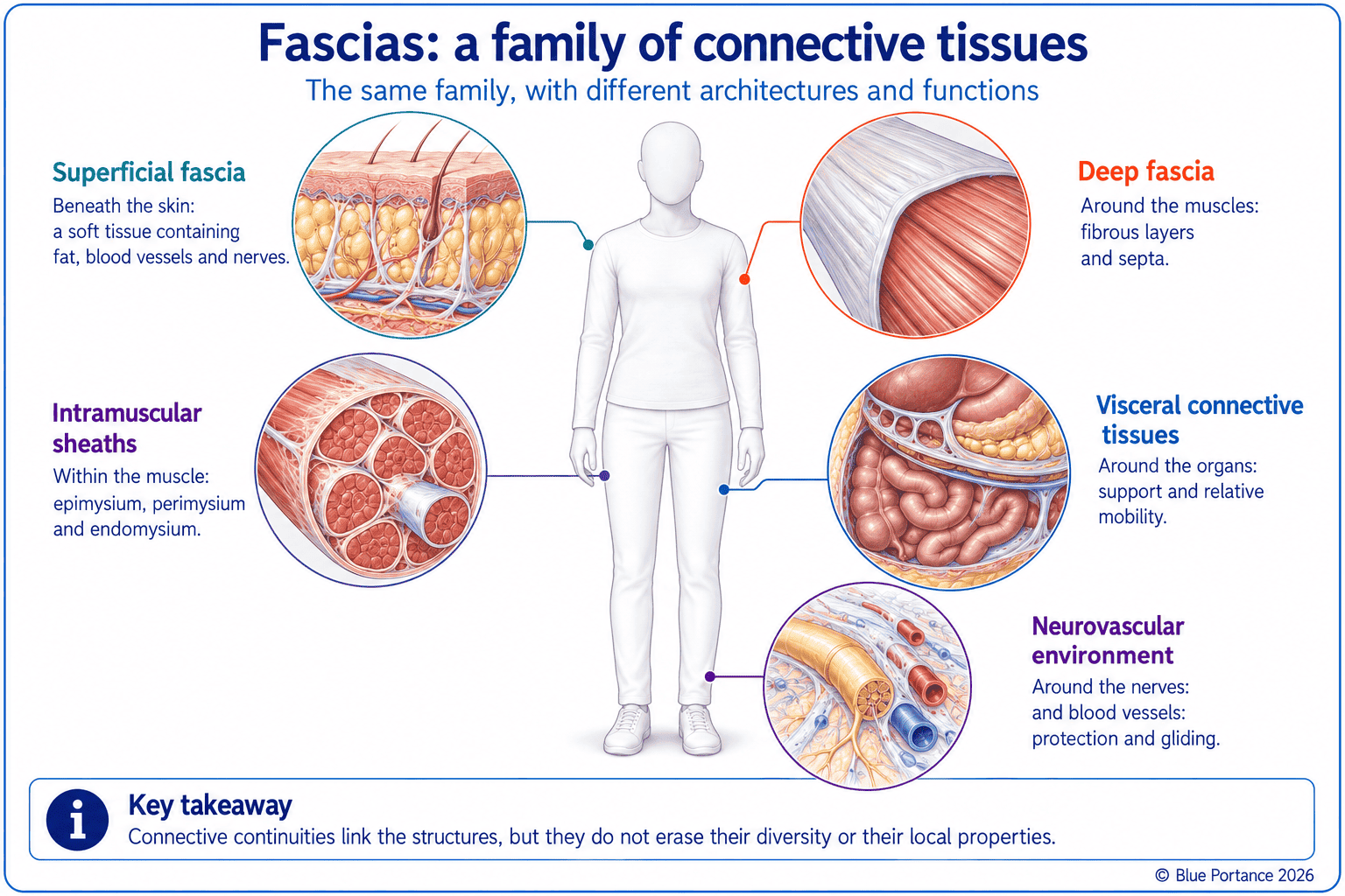
Summary — Fascia forms a family of connective tissues that wrap, separate, connect, and accompany the structures of the body. These tissues combine cells with an extracellular matrix composed notably of fibers, a highly hydrated ground substance, and adhesion molecules. Their architecture varies from region to region: superficial fascia, deep fascia, muscle sheaths, visceral planes, and neurovascular interfaces. This diversity thus enables them to provide support and relative mobility at the same time.
1. One Word for Different Tissues
The term fascia long referred to the connective sheets visible during dissection. In particular, its contemporary usage can include sheaths, septa, aponeuroses, retinacula, and certain supporting tissues. It is therefore more accurate to speak of fascial tissues, in the plural, than of a single uniform fascia (Adstrum et al., 2017). Synthesis works written for a wider readership than anatomists alone have also relayed this shift. This broader notion is sometimes referred to as the “fascial system” (Lesondak, 2019). Admittedly, this popularization remains useful for raising awareness of the importance of these tissues. However, it can foster the image of a continuous, uniform web—an image this chapter specifically sets out to qualify.
One Family, Different Architectures
Thus, a supple subcutaneous layer, a dense aponeurosis, and a visceral sheath belong to the same broad family. Yet their composition, thickness, and fiber orientation differ. In fact, function cannot be inferred from the name “fascia” alone.
This diversity is directly reflected in anatomical nomenclature. For example, the fascia lata wraps the thigh and continues laterally as the iliotibial band. This band, reinforced by dense, parallel fibers, is oriented to resist repeated tensile loading along a preferred axis. By contrast, the thoracolumbar fascia is a multilayered sheet. It connects the deep trunk muscles to the superficial muscles of the back and limbs, and its fiber orientation changes from one layer to the next. As for the plantar fascia, it forms a thick fibrous band beneath the arch of the foot. Its tension varies with posture and weight-bearing. Three structures, three architectures, three distinct mechanical roles—all designated by the same word.
2. An Organized Extracellular Matrix
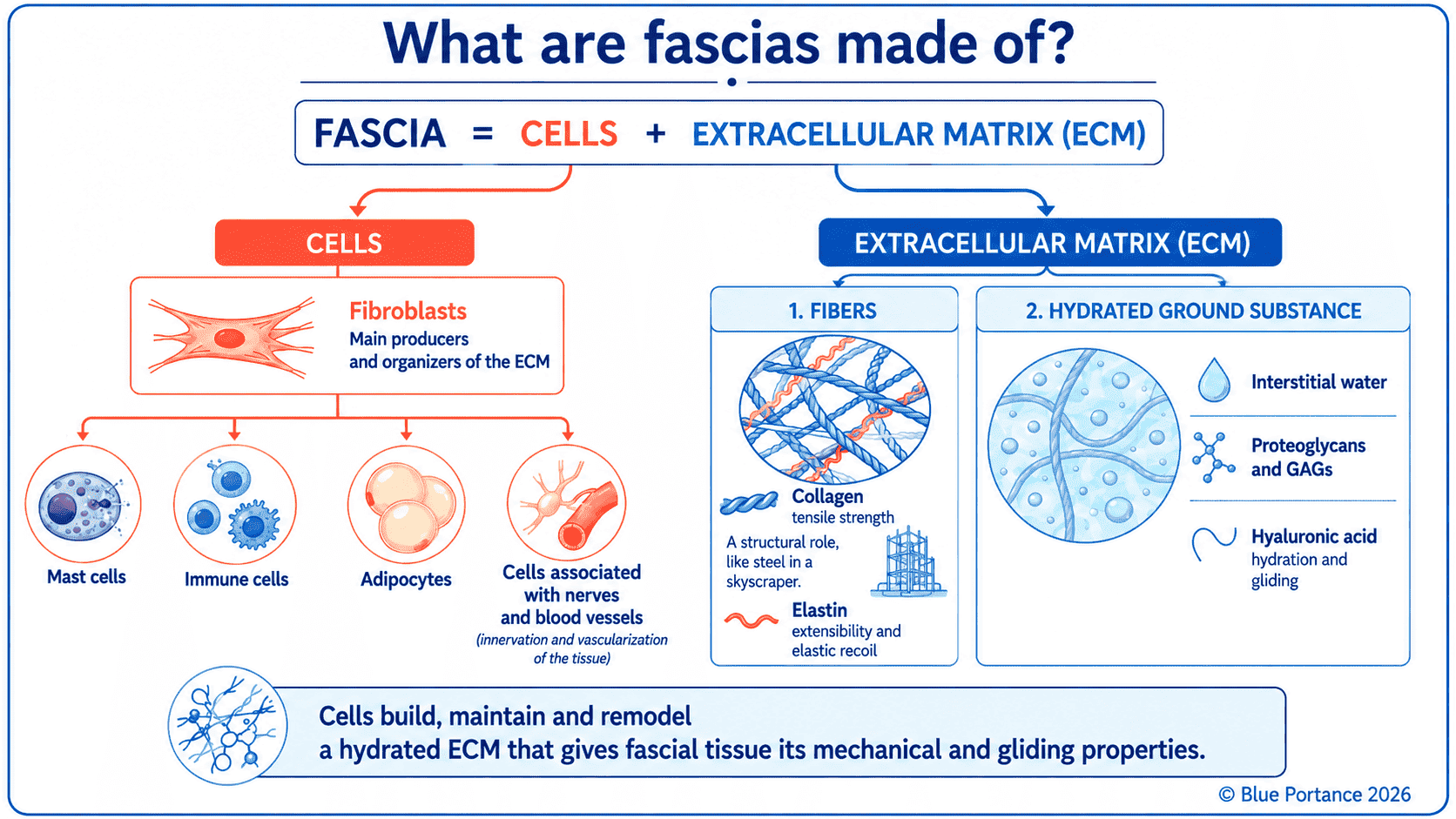
Fascia combines cells—mainly fibroblasts—with an extracellular matrix that these cells synthesize and remodel. Notably, this matrix comprises two major, complementary components: fibers and the highly hydrated ground substance that surrounds them. Their proportions and organization vary considerably from one tissue to another. A supple, hydrated superficial fascia thus does not have the same composition as a tendon or a dense aponeurosis (Lodish et al., 2000).
Collagen: The Mechanical Framework
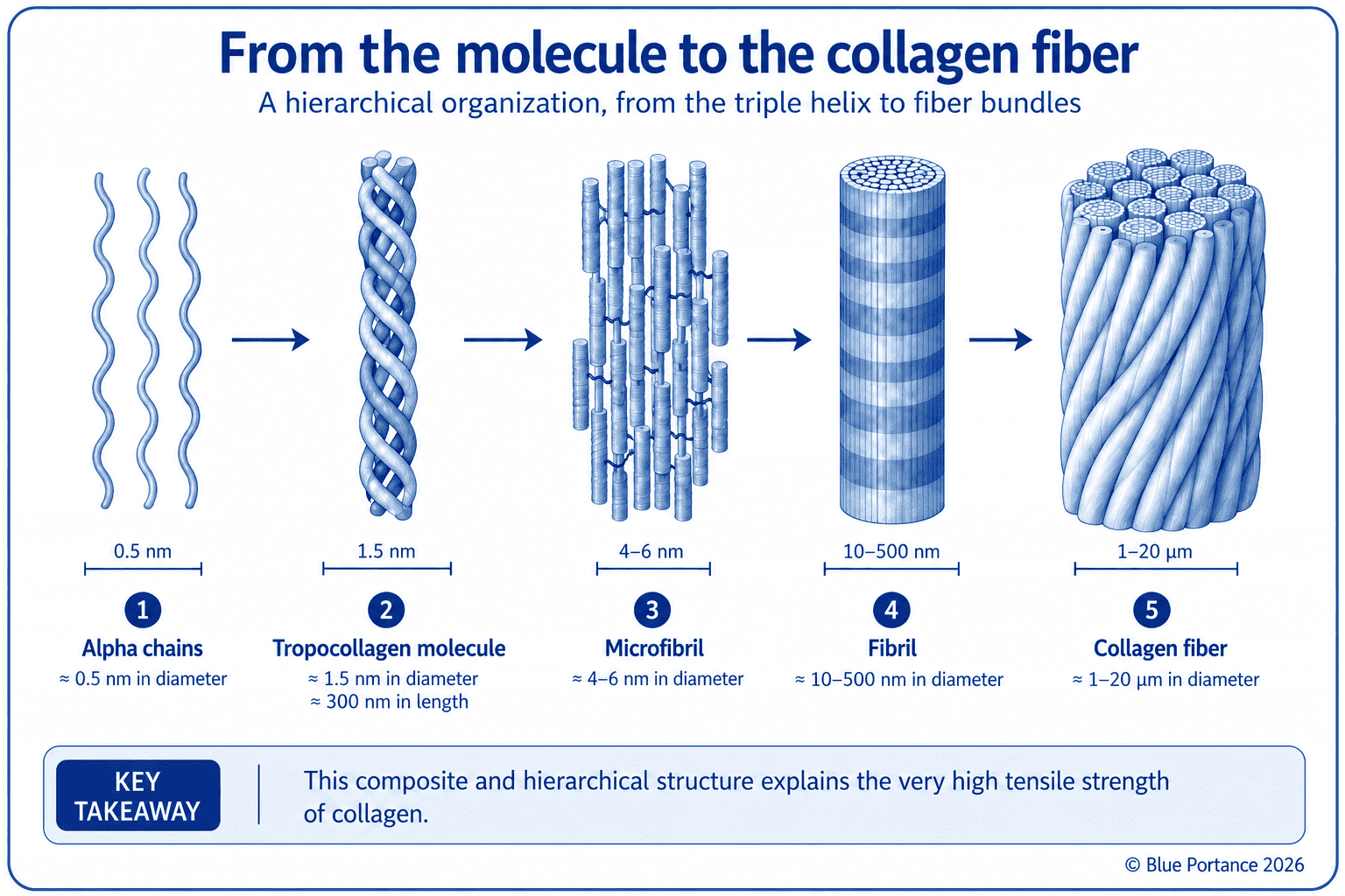
Collagen fibers form the mechanical framework of fascia. In fact, collagen is the most abundant protein in the human body. Each α chain is a polypeptide chain made up of a sequence of amino acids. Three α chains then coil into a triple helix to form a collagen molecule. These molecules first align in parallel, with a periodic longitudinal offset, into microfibrils. These form units of supramolecular organization, roughly 4 to 6 nm in diameter, without necessarily constituting a distinct morphological level in every description (Holmes & Kadler, 2006). At the fibril scale, this same periodic offset creates alternating gap and overlap zones. This alternation gives rise to the fibrils’ characteristic banding, with a D-period of about 67 nm (Gautieri et al., 2011).
The fibrils formed in this way then assemble into fibers, and then into oriented bundles. Their diameter varies considerably, on the order of 10 to 500 nm depending on the tissue, age, and observation method (Zhang et al., 2021).
An Architecture That Explains Tensile Strength
Thus, this hierarchical architecture, reinforced by the bonds between molecules, provides exceptional tensile strength. At the molecular scale, some estimates place the specific strength of collagen at a level comparable to, or even higher than, that of steel. Collagen can indeed tolerate much greater strain (Buehler, 2006). This performance does not, however, translate directly to an entire fascia, whose behavior also depends on its architecture and hydration. These fibers notably enable tendons, ligaments, aponeuroses, and deep fascia to withstand large forces without rupturing.
Elastic Fibers and Ground Substance
Elastic fibers allow stretching and a return toward the original shape. Composed notably of elastin and microfibrils, they are much less abundant than collagen in most fascial tissues. Overall, their proportion depends on the location and function of the tissue. They provide a capacity for reversible deformation, without on their own accounting for all of fascia’s elasticity.
The ground substance is the fluid medium of the matrix. Amorphous, transparent, and variable in consistency, it fills the space between cells and fibers. Being highly hydrated, it takes part in the exchange of nutrients, signaling molecules, and waste products between capillaries, lymph, and cells. It thus forms the immediate environment of each cell. Its consistency can be relatively fluid in loose connective tissue and much firmer in a specialized tissue such as cartilage.
This ground substance contains, notably, proteoglycans, glycosaminoglycans, and hyaluronan (hyaluronic acid). Through their strong affinity for water, proteoglycans and glycosaminoglycans contribute to the hydration of the matrix. They also contribute to its gel-like consistency and to its resistance to compressive forces. Finally, hyaluronan contributes to viscosity and to the lubrication of interfaces. It thus promotes gliding between fibers, bundles, and fascial layers (Cowman et al., 2015). It is not simply an oil: its properties depend notably on its concentration, its temperature, and its molecular interactions.
A Matrix Connected to the Cells
The matrix and the cells are mechanically connected. Transmembrane adhesion receptors, notably integrins, link the extracellular matrix to the cytoskeleton. They take part in detecting mechanical loading such as tension, compression, or shear. They also convert it into biochemical signals: this is mechanotransduction (Ingber, 2003). Chapter 6 will detail how it works and its effects on tissue remodeling.
3. Complementary Anatomical Families
In fact, the superficial fascia connects the skin to the deeper planes and contains fat, vessels, and nerves. Depending on the nomenclature adopted, the term can refer to the entire stratified subcutaneous tissue or, more strictly, to its deep membranous connective layer (Benjamin, 2009). In either case, it is not homogeneous throughout its depth. It classically comprises a superficial fatty layer and a deeper, more fibrous and membranous layer, separated by a gliding plane. This two-layer organization explains why certain procedures (palpation, surgery, manual manipulation) encounter different resistance depending on the depth reached.
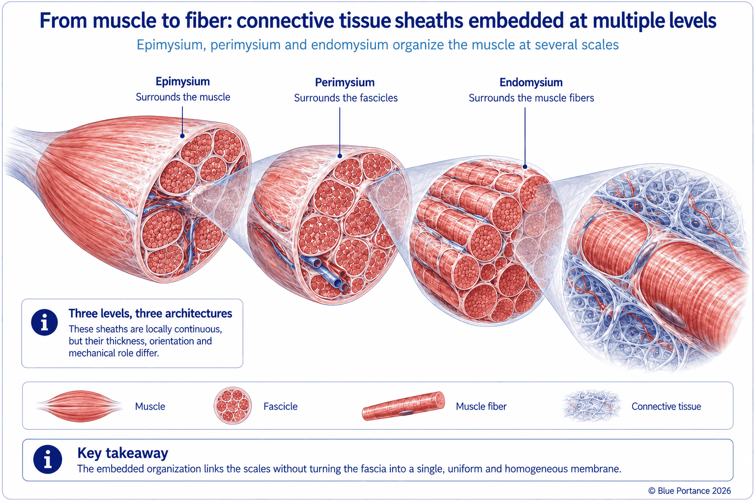
Furthermore, the deep fascia wraps the muscles, delimits compartments, and forms attachment zones. It becomes locally specialized according to the stresses it bears. Around joints, it thickens into retinacula that hold the tendons against the bone during movement. Between two muscles or muscle groups, it forms septa that separate muscle compartments. These septa also serve as anchoring surfaces for muscle fibers. Aponeuroses, for their part, are broad, flattened tendinous structures associated with certain muscles. They distribute forces over a wide area rather than concentrating them at a single point (Kumka & Bonar, 2012). Within the muscle itself, the endomysium, perimysium, and epimysium organize the fibers at several scales, from the individual muscle fiber to the whole muscle.
Visceral Tissues and the Neurovascular Environment
Likewise, the visceral connective tissues follow a comparable logic, on a different scale. Connective folds (mesenteries, visceral ligaments), for example, hold the abdominal organs in place and carry their blood supply. They thus connect the organs to the abdominal wall while allowing changes in their volume and letting them glide against one another. They also allow the movements related to breathing, posture, or digestive filling (Stecco, 2015).
Around nerves, within a broad functional understanding of the fascial system, a layered, nested organization follows the same principle. The endoneurium surrounds each nerve fiber, the perineurium encloses bundles of fibers, and the epineurium wraps the entire nerve. At the scale of the peripheral nervous system, this architecture mirrors that of the endomysium, perimysium, and epimysium in muscle. In both cases, these sheaths protect the structure they surround while preserving its mobility relative to neighboring tissues.
4. Continuity Does Not Mean Uniformity
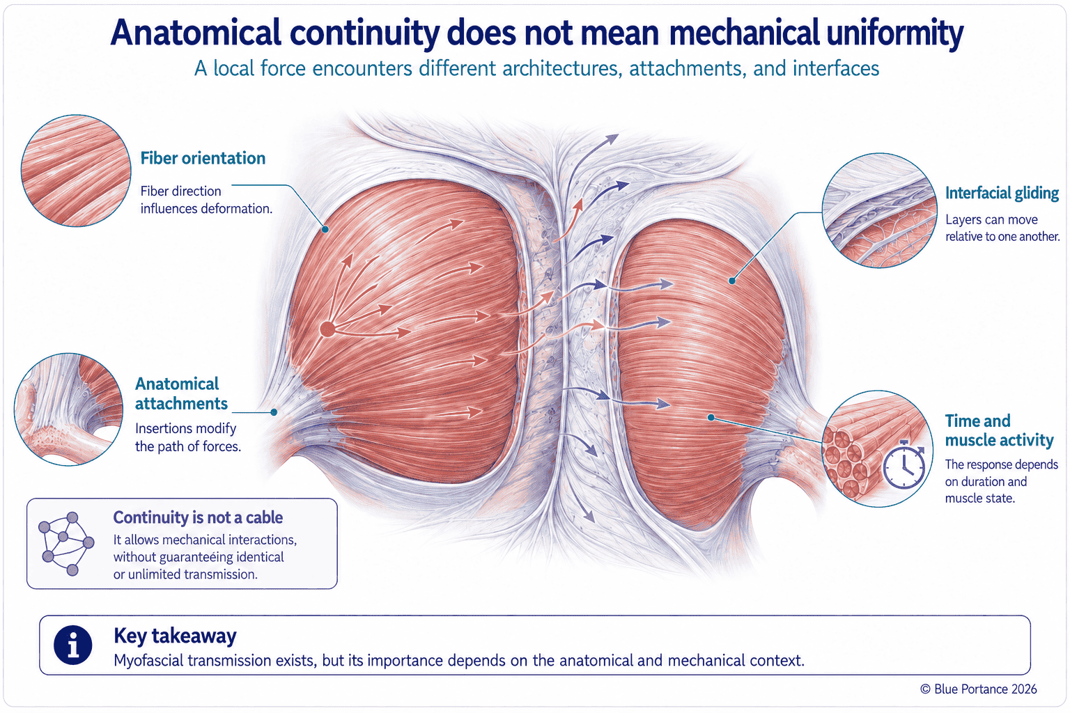
In fact, anatomical continuities allow mechanical interactions between neighboring structures. They do not prove that a local force is transmitted to the whole body without attenuation. The response depends on five factors: geometry, attachments, fiber orientation, muscle activity, and time. Together, these factors determine whether a stress remains localized or spreads (Huijing, 2003).
A contrast helps illustrate these factors. Thus, a tendon, a dense structure with parallel fibers, efficiently transmits the force of a muscle to the bone. Its geometry and fiber orientation are specifically organized for this purpose. By contrast, a loose connective continuity between two neighboring compartments does not have this function. It allows gliding and mutual adaptation, not efficient transfer of tension. Two structures described as “fascia” can therefore play opposite mechanical roles depending on their architecture. Moreover, a connective tissue’s response to loading is neither instantaneous nor fixed. It also depends on how long the force is applied and on the prior state of the tissue—which the next chapter explores in more depth.
Tensegrity can help represent a stability achieved through a balance between tension and compression. At the whole-body scale, however, it remains an interpretive model, not a law sufficient to explain every movement or every pain.
5. A Living Tissue
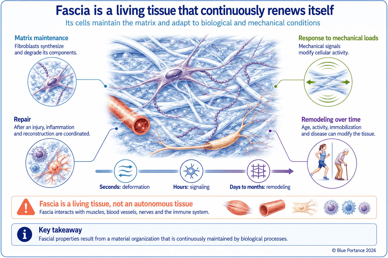
Indeed, fascia contains active cells, variable vascularization, and nerve endings. It renews and remodels itself with age, activity, immobilization, injury, and inflammation. Saying that fascia is living does not mean that it acts autonomously, but that its properties depend on biological regulation.
This renewal takes place at the cellular scale. Fibroblasts do not simply produce the extracellular matrix once and for all. They continuously break it down and resynthesize it. This balance between synthesis and degradation can shift in one direction or the other depending on the loading the tissue receives. Appropriate mechanical loading can promote the renewal of the matrix and its organization along lines of force. Conversely, prolonged immobilization leads to changes in its composition and organization. The response, however, depends on the tissue and on the intensity and duration of the load (Chiquet et al., 2009). It is this same principle of biological regulation, rather than a fixed mechanical property, that is at play here. It explains why fascia can stiffen or regain suppleness depending on how it is loaded over time.
Furthermore, vascularization and innervation are not uniform either. Some fasciae are richly vascularized and innervated, others far less so, which locally modulates their healing capacity and their sensitivity (Stecco, 2015). Chapter 7, devoted to the neurosensory interface of fascia, will examine this fascial innervation in more detail, particularly its sensory component.
Key Takeaways
- In short, fascia is a family of connective tissues, not a single membrane.
- Its organization thus combines cells with an extracellular matrix made of fibers and a highly hydrated ground substance.
- Collagen forms a framework that is highly resistant to tension; elastic fibers, for their part, contribute to reversible deformation.
- The ground substance, in turn, also contributes to exchanges, to resistance to compression, and to gliding between structures.
- Anatomical continuities therefore allow forces to be transmitted, but do not imply unlimited propagation.
Frequently Asked Questions
Does fascia surround only the muscles?
Do all types of fascia have the same properties?
Does fascia form a single web?
What is the difference between fascia, a tendon, and a ligament?
Scientific References
Cited References (1/2)
- Adstrum S, Hedley G, Schleip R, Stecco C, Yucesoy CA. Defining the fascial system. J Bodyw Mov Ther. 2017;21(1):173–177.
- Benjamin M. The fascia of the limbs and back—a review. J Anat. 2009;214(1):1–18.
- Buehler MJ. Nature designs tough collagen: explaining the nanostructure of collagen fibrils. Proc Natl Acad Sci USA. 2006;103(33):12285–12290.
- Chiquet M, Gelman L, Lutz R, Maier S. From mechanotransduction to extracellular matrix gene expression. Biochim Biophys Acta. 2009;1793:911–920.
- Cowman MK, Lee HG, Schwertfeger KL, McCarthy JB, Turley EA. The content and size of hyaluronan in biological fluids and tissues. Front Immunol. 2015;6:261.
- Gautieri A, Vesentini S, Redaelli A, Buehler MJ. Hierarchical structure and nanomechanics of collagen microfibrils from the atomistic scale up. Nano Lett. 2011;11(2):757–766.
- Holmes DF, Kadler KE. The 10+4 microfibril structure of thin cartilage fibrils. Proc Natl Acad Sci U S A. 2006;103(46):17249–17254.
Cited References (2/2)
- Huijing PA. Muscular force transmission necessitates a multilevel integrative approach. Exerc Sport Sci Rev. 2003;31(4):167–175.
- Ingber DE. Tensegrity I. Cell structure and hierarchical systems biology. J Cell Sci. 2003;116:1157–1173.
- Kumka M, Bonar J. Fascia: a morphological description and classification system. J Can Chiropr Assoc. 2012;56(3):179–191.
- Lesondak D. Le Fascia. [Fascia.] Éditions Ressources Primordiales; 2019.
- Lodish H, Berk A, Zipursky SL, et al. Molecular Cell Biology. 4th ed. W. H. Freeman; 2000.
- Stecco C. Functional Atlas of the Human Fascial System. Elsevier; 2015.
- Zhang S, Ju W, Chen X, Zhao Y, Feng L, Yin Z, Chen X. Hierarchical ultrastructure: an overview of what is known about tendons and future perspective for tendon engineering. Bioact Mater. 2021;8:124–139.
Back to the Fascia guide
Next chapter: How Does Fascia Behave Under Stress? →
Note: this content explains general mechanisms. It does not constitute a medical diagnosis or a therapeutic recommendation.
© Gil Ayache. The original concepts, biomechanical models, diagrams, terminology, graphic representations, foundational figures, original texts, and principles presented on this page are works protected by copyright. They are made available to Blue Portance under an intellectual property license agreement, without transfer of economic rights or of authorship.
