


1. The central question
At this stage, one question becomes central — and it determines absolutely everything else:
why does this coccyx hurt?
Is it because it is structurally damaged?
Or because it is functionally trapped in an environment that no longer allows it to play its role?
This distinction may seem theoretical. In reality it is fundamental, because it determines:
- which examinations are useful,
- the logic of care,
- and above all, the reasons why some treatments fail while others work.
2. Structural causes: when the bone is involved
In some cases of coccydynia, the pain is linked to an objectifiable abnormality of the coccyx itself.
The main findings are:
- an anterior or posterior dislocation (Postacchini & Massobrio, 1983; Maigne et al., 2000),
- excessive hypermobility (Maigne & Tamalet, 1996; Lirette et al., 2014),
- more rarely, a coccygeal spur (spicule) (Fogel et al., 2004; Lirette et al., 2014).
These abnormalities share an essential feature:
they are often invisible on a standard radiograph taken standing or lying down.
It is precisely this observation that led Maigne and Tamalet to develop a dynamic sitting/standing radiograph protocol, now considered the reference examination for investigating coccydynia (Maigne & Tamalet, 1996).
What this dynamic imaging shows is not only the shape of the coccyx, but its behavior under load (Maigne & Tamalet, 1996; Lirette et al., 2014).
A coccyx can look perfectly normal at rest…
Yet it can dislocate, shift, or become hypermobile as soon as the person sits down (Maigne & Tamalet, 1996).
This is often the moment when the diagnosis shifts:
the pain is no longer “unexplained”; it becomes mechanically coherent.
3. When imaging is normal… but the pain is very real
However, in a significant number of cases, imaging examinations — even dynamic ones — reveal no obvious structural abnormality (Fogel et al., 2004; Lirette et al., 2014).
And yet, the pain is there.
Intense. Disabling.
This is where the notion of functional coccydynia comes in (Fogel et al., 2004).
In these situations, the coccyx is not damaged, but it is trapped in an environment that has lost its capacity to adapt:
- pelvic floor hypertonia (Vodusek, 2004),
- chronic muscle tension (Fogel et al., 2004),
- loss of overall pelvic mobility,
- repeated postural stress.
A muscular environment that traps the coccyx
The coccyx then becomes the fixation point of a broader imbalance.
In reality, it is not the bone that is diseased;
it is the system around it that can no longer absorb the stress.
If imaging remains silent, the key often lies in the dynamics of the soft tissues that act as “guy wires” for the coccyx.
In some patients, functional coccydynia may reflect pelvic floor hypertonia.
The levator ani and the coccygeus muscle attach directly to the lateral borders of the coccyx (Vodusek, 2004).
Under chronic stress, or as a protective reflex after an impact, these muscles can enter a cycle of permanent contracture (Fogel et al., 2004).
In some patients, the coccyx is then “pinned” by these overly tight muscular guy wires, losing its ability to tilt when moving into the seated position (Fogel et al., 2004).
As a result, during the clinical examination, reproducing the pain by putting these muscles under tension supports the hypothesis that the coccyx is being mechanically stressed by an overly rigid myofascial environment (Fogel et al., 2004).
This mechanism explains why some people show no abnormality on Maigne’s dynamic radiographs, yet suffer from a real mechanical stress imposed by their own musculature (Maigne & Tamalet, 1996; Fogel et al., 2004).
Figure 5 — The pelvic muscular sling and the coccyx
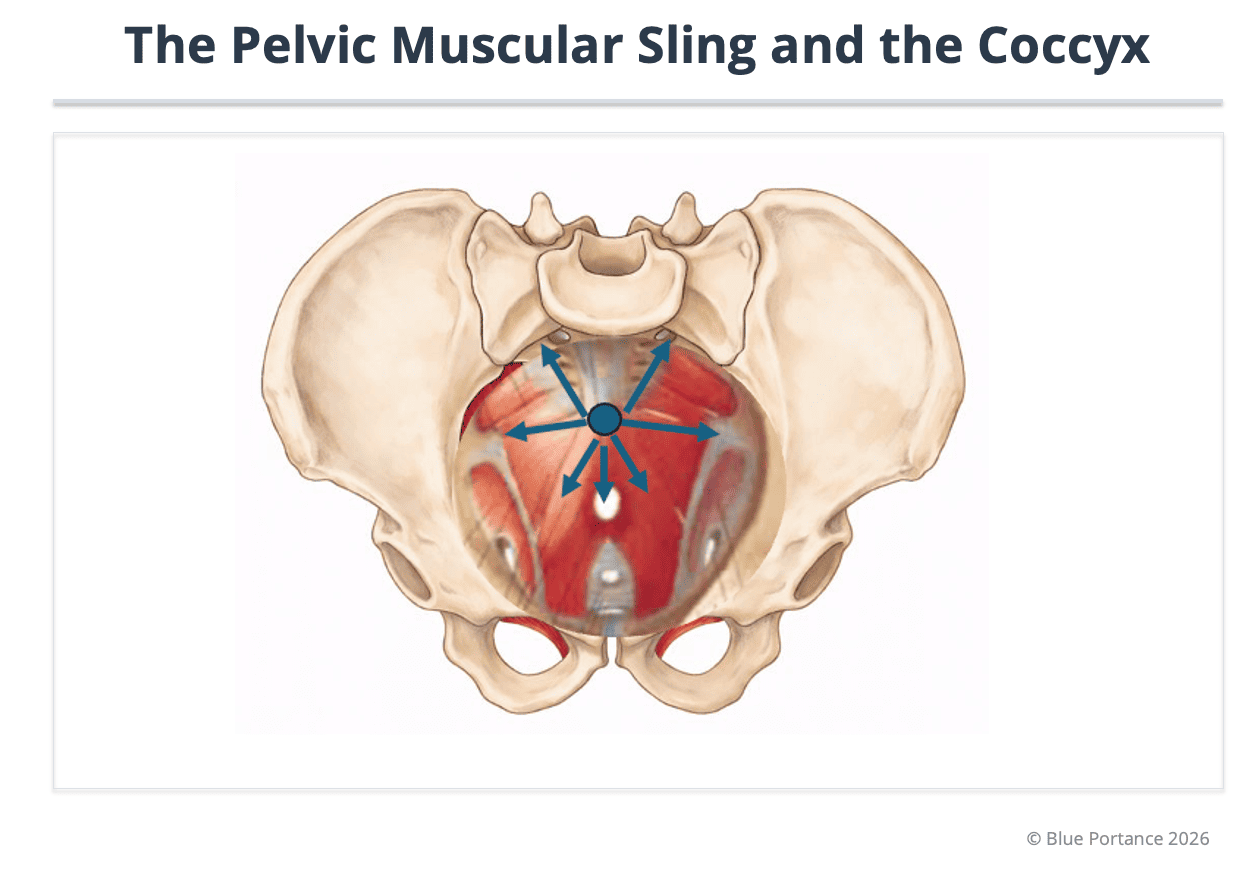
Caption: In short, this figure shows how the attachment of the levator muscles to the borders of the coccyx creates tension that can “imprison” it and block its physiological mobility (Vodusek, 2004; Fogel et al., 2004).
4. The key role of the clinical examination
It is in these functional forms that the clinical examination takes on its full value.
Palpation makes it possible to:
- reproduce the pain,
- assess the mobility of the coccyx,
- identify areas of muscle tension,
- detect protective reactions of the pelvic floor.
A coccyx that is painful on palpation, without any radiological abnormality, is not a “false problem.”
It often reflects local hypersensitization or an indirect mechanical stress (Fogel et al., 2004).
Here again, the pain makes sense.
It points instead to a loss of functional freedom, not a visible lesion.
The “structural vs functional” distinction does not set “real” pain against “imaginary” pain. It contrasts an objectifiable bony lesion or instability with an indirect mechanical stress produced by the myofascial environment and postural dynamics.
When imaging is normal, the functional hypothesis becomes central: it requires looking for what acts as a “guy wire” on the coccyx (pelvic floor, tension, loss of mobility) and for what prevents the system from absorbing stress in the seated position (Vodusek, 2004; Fogel et al., 2004).
In other words: the bone may be intact, but the system around it may have become too rigid to remain compatible with the load of sitting (Maigne & Tamalet, 1996; Lirette et al., 2014).
5. A structuring metaphor: a healthy part in a seized mechanism
This situation can be compared to a perfectly intact mechanical part … built into a seized-up system.
- The part (the coccyx) is healthy.
- The axes around it (muscles, ligaments, posture) are rigid.
- Overall movement becomes constrained.
- Stress concentrates on the most exposed point.
The pain is not proof of an intrinsic fragility.
It is the signal of an imbalance in how stress is distributed.
6. Why this distinction prevents so many errors
Failing to distinguish structural from functional leads to two frequent dead ends:
- searching endlessly for a visible lesion that does not exist,
- or, conversely, treating a very real mechanical instability “functionally.”
In both cases, the person becomes exhausted, starts to doubt, and ends up believing the pain is “all in their head.”
Yet, whether structural or functional, coccydynia is never imaginary.
It is always the expression of a system that can no longer adapt.
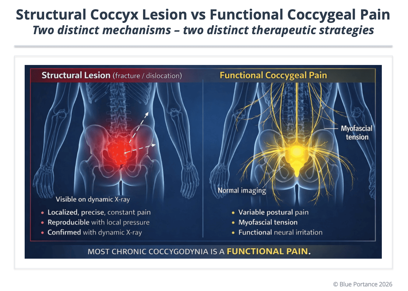
Purpose: to show why static imaging can be normal while pain that is mechanically coherent persists, and why the clinical examination and functional reading remain decisive (Maigne & Tamalet, 1996).
Scientific references
- Maigne, J. Y., & Tamalet, B. (1996). Standardized radiologic protocol for the study of common coccygodynia and characteristics of the lesions observed in the sitting position. Spine, 21(22), 2588–2593.
- Fogel, G. R., Cunningham, P. Y., & Esses, S. I. (2004). Coccygodynia: Evaluation and management. Journal of the American Academy of Orthopaedic Surgeons, 12(1), 49–54.
- Vodušek, D. B. (2004). Anatomy and neurocontrol of the pelvic floor. Digestion, 69(2), 87–92.
- Postacchini, F., & Massobrio, M. (1983). Idiopathic coccygodynia. Journal of Bone and Joint Surgery, 65(8), 1116–1124.
- Maigne, J. Y., Doursounian, L., & Chatellier, G. (2000). Causes and mechanisms of common coccydynia: role of body mass index and coccygeal trauma. Spine, 25(23), 3072–3079.
- Lirette, L. S., Chaiban, G., Tolba, R., & Eissa, H. (2014). Coccydynia: An overview of the anatomy, etiology, and treatment of coccyx pain. Ochsner Journal, 14(1), 84–87.
© Gil Ayache. The concepts, diagrams, terminology, and principles presented on this page constitute a work protected by copyright. They are made available to Blue Portance under an intellectual property license agreement, without transfer of economic rights or of authorship.
