


1. Mechanical causes: where does it really get stuck?
When the coccyx becomes painful, it is tempting to look for a single cause: a fracture, a dislocation, a “displaced bone.”
The coccyx is a terminal point.
It sits at the end of a long, heavily loaded mechanical chain: the spine and the pelvis.
It therefore receives part of the stress generated by:
- body weight,
- prolonged sitting,
- micro-trauma,
- pelvic mobility,
- the condition of the soft tissues,
- musculoligamentous attachments.
From this perspective, “mechanical” coccydynia does not refer only to a bony lesion, but to a failure of the coccygeal segment to adapt to load and movement. Clinical series and dynamic imaging show that mobility abnormalities can be present even when standard imaging looks “normal” [1] (Maigne et al., 2000).
2. Falls, impacts, and forgotten trauma
In general, the most classic cause of coccydynia is direct trauma: a fall onto the buttocks, an impact (sports, a slip, an accident), or a blow to the coccyx.
This trauma can:
- cause a fracture (less common),
- lead to a dislocation of the coccyx,
- subtly alter its mobility without any obvious lesion.
An important point is that the initial trauma may be old, sometimes forgotten, and that the pain may appear or worsen later. This delayed onset, frequently reported, is explained by the gradual accumulation of stress (sitting, posterior pelvic tilt, micro-trauma) until a threshold of functional tolerance is exceeded [1] (Maigne et al., 2000).
The coccyx can therefore remain “off-center” or unstable long after the triggering event, and become painful when the cumulative mechanical stress exceeds a tolerance threshold.
3. Childbirth: a specific mechanical stress
In women, childbirth is a particular mechanical context. To allow the baby to pass, the coccyx must move backward.
This mechanism can induce:
- a dislocation,
- residual hypermobility,
- or persistent pain related to a loss of local stability [1] (Maigne et al., 2000).
The nuance is essential: childbirth does not systematically cause a dislocation, but it can represent a maximal stress on a sacrococcygeal joint that is already vulnerable, with imbalanced mobility and secondary tissue irritability [1] (Maigne et al., 2000).
This is one of the reasons why coccydynia is markedly more common in women.
4. Prolonged sitting: the silent micro-trauma
However, not all cases of coccydynia begin with a clear-cut impact. In some people, the pain sets in gradually.
Sitting for a long time on a hard or poorly suited surface concentrates pressure:
- on the ischial tuberosities,
- but also on the tip of the coccyx, especially when the pelvis tilts backward.
Through repetition, these micro-stresses can:
- irritate the soft tissues,
- disturb coccygeal mobility,
- promote the onset of chronic pain [2] (Lirette et al., 2014).
It is the mechanical equivalent of a door that never slams outright, but forces a little… every time it opens.
“Beyond the duration of sitting, it is the very shape of the contact between the seat and the coccyx that determines the areas of stress.”
Imaging research has refined our understanding of risk according to the morphology and dynamics of the coccyx.
Changes in our lifestyles have also introduced the “screen posture”: a posteriorly tilted pelvis, a rounded spine, and increased pressure on the tip of the coccyx.
5. Mechanical abnormalities
Instability, hypermobility, spicule: when the mechanics go awry
Reference studies show that coccydynia is frequently associated with objectifiable mechanical abnormalities… provided they are looked for properly [1,3] (Maigne et al., 2000; Nathan et al., 2010).
-
Anterior or posterior dislocation of the coccyx
An abnormal displacement relative to the sacrum, typically revealed by dynamic sitting/standing radiographs [1] (Maigne et al., 2000). -
Coccygeal hypermobility
Excessive mobility that makes sitting unstable and irritating — a configuration described in clinical series [1] (Maigne et al., 2000). -
Coccygeal spur (bony spicule)
A terminal outgrowth likely to irritate the soft tissues, particularly during prolonged weight-bearing [3] (Nathan et al., 2010).
These abnormalities are often invisible on a standard radiograph. They sometimes require a dynamic sitting/standing radiograph, or even more targeted examinations depending on the context [1,2] (Maigne et al., 2000; Lirette et al., 2014).
“Everything is normal on the X-rays.”
In reality, this statement does not rule out a mechanical cause: it frequently indicates that the examination did not explore functional mobility.
The pain may stem from dynamic instability, localized overload, and a loss of adaptability
that only show up under weight-bearing [1,3] (Maigne et al., 2000; Nathan et al., 2010).
6. Factors that perpetuate and aggravate the pain
Even when a mechanical abnormality exists, certain factors act as amplifiers.
One of the best documented is a high body mass index. Maigne et al. showed a clear association between coccydynia and excess weight [1] (Maigne et al., 2000).
While weight is an obvious load factor, the very nature of the contact surface and the geometry of the seat can turn moderate stress into focused stress.
Obesity alters pelvic kinematics during sitting: in people with a high BMI, posterior pelvic tilt is often more pronounced, which increases pressure on the tip of the coccyx [1] (Maigne et al., 2000).
In addition, recent reviews emphasize that prolonged sitting acts not only through pressure, but also through local tissue irritation and the potentiation of chronicity mechanisms [2] (Lirette et al., 2014).
Added to this are:
- postural imbalances of the pelvis,
- a loss of overall mobility,
- chronic muscle tension in the pelvic floor.
The coccyx then becomes the weak link in a system that no longer distributes stress correctly.
Mechanical coccydynia can be compared to a poorly adjusted door hinge.
- The door (the pelvis) is heavy.
- The hinge (the coccyx) is small.
- As long as the hinge works within its tolerance zone, all is well.
- But as soon as it is bent, locked, or too loose, every opening becomes difficult… and eventually painful.
In reality, the pain is not the problem in itself.
It is the alarm signal of a mechanism that has lost its capacity to adapt.
Figure — Mechanical causes of coccydynia
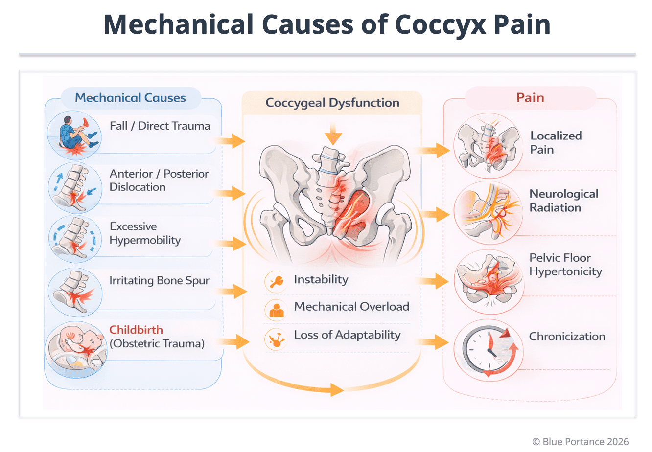
Doctrinal explanation: the figure does not propose a “single cause,” but a sequence. The same case of coccydynia may combine an initial stress (a fall or childbirth), a change in mobility (dislocation or hypermobility), and then repeated overload while sitting. The clinical consequence is not only localized pain: depending on the individual and the tissues involved, it may be accompanied by radiating pain and protective hypertonia of the pelvic floor, contributing to chronicity [2] (Lirette et al., 2014).
This diagram is consistent with the central idea highlighted in dynamic studies: what standard imaging sometimes calls “normal” may correspond to a dysfunction under load, visible only when the coccyx is studied in a functional situation [1] (Maigne et al., 2000).
Scientific references
- Maigne, J. Y., Doursounian, L., & Chatellier, G. (2000). Causes and mechanisms of common coccydynia: Role of body mass index and coccygeal trauma. Spine, 25(23), 3072–3079.
- Lirette, L. S., Chaiban, G., Tolba, R., & Eissa, H. (2014). Coccydynia: An overview of the anatomy, etiology, and treatment of coccyx pain. Ochsner Journal, 14(1), 84–87.
- Nathan, S. T., Fisher, B. E., & Roberts, C. S. (2010). Coccydynia: A review of pathoanatomy, aetiology, treatment, and outcome. Journal of Bone and Joint Surgery (British Volume), 92-B(12), 1622–1627.
© Gil Ayache. The concepts, diagrams, terminology, and principles presented on this page constitute a work protected by copyright. They are made available to Blue Portance under an intellectual property license agreement, without transfer of economic rights or of authorship.
