


Introduction — “It feels like I’m sitting on a nail”
“When I sit down, it’s still okay.
But after a few minutes, a deep pain sets in, as if something were pushing inside.
And when I stand up, it’s like a lightning bolt.”
This kind of description comes up with surprising consistency among people suffering from coccydynia. Indeed, the pain is rarely diffuse. It is precise, centered, often described as a nail, a dagger, sometimes a dull burning, located at the lower end of the spine (Nathan, Fisher & Roberts, 2010).
Gradually, sitting becomes uncomfortable, then outright painful. The body develops avoidance strategies: sitting on one side, leaning the trunk forward, constantly changing position, standing up “before it hurts too much.” Simple activities — working at a screen, driving, watching a movie — thus turn into permanent burdens (Nathan, Fisher & Roberts, 2010).
Despite this very specific symptom pattern, coccydynia is still frequently misidentified. It is confused with low back pain, sciatica, or anoperineal pain, or wrongly classified as “functional” because no abnormality is visible on imaging. This confusion is not trivial: it leads to inappropriate care, prolonged diagnostic wandering, and the mistaken idea that “nothing explains the pain” (Nathan, Fisher & Roberts, 2010).
The reality, however, is different. In most cases, coccydynia rests on identifiable anatomical, biomechanical, and neuro-functional mechanisms, provided one looks in the right place and with the right interpretive framework. That is precisely the purpose of this page (Nathan, Fisher & Roberts, 2010).
1. The coccyx: a small bone… with a major functional impact
1.1 A discreet bone, but structurally strategic
The coccyx is located at the lower end of the spine. It consists of three to five vertebral segments, more or less fused (Woon & Stringer, 2012). Given its small size, its limited mobility, and how little it is understood, it is sometimes perceived as a secondary, even useless, bone. In reality, this perception is wrong (Standring, 2021).
Anatomically and functionally, the coccyx occupies a strategic position:
- it is a lower anchor point for major pelvic floor muscles (Standring, 2021; Vodušek, 2004),
- it contributes to the stability of the pelvic ring (Standring, 2021),
- finally, it lies at the direct interface between the spine, the pelvis, and the seating surface (Woon & Stringer, 2012).
In other words, the coccyx is not a mere terminal segment: it is an element of mechanical cohesion between structures subjected to substantial stress, particularly in the seated position (Nathan, Fisher & Roberts, 2010).
2. The coccyx as a support point for the pelvic floor
2.1 A fundamental muscular crossroads
Figure 1 highlights a central point: the coccyx is a lower convergence point of the pelvic floor. Several essential muscle bundles are organized around it, in particular the posterior components of the levator ani and the coccygeus muscle, together with the associated ligamentous structures (Standring, 2021; Vodušek, 2004).
2.2 Cohesion, stability, and support
These attachments give the coccyx the role of an active support. It does not merely receive forces: it enables the coordinated tensioning of the pelvic sling, ensuring the suspension and cohesion of the whole. Despite its small size, the coccyx contributes to the overall stability of the pelvis, to the distribution of muscular tension, and to the functional continuity between the spine and the pelvic floor (Standring, 2021; Vodušek, 2004).
When this support point is functional, the system remains stable and silent. When its position, mobility, or integrity is altered, the pelvic sling can become disorganized, increasing local stress and making pain more likely (Nathan, Fisher & Roberts, 2010).
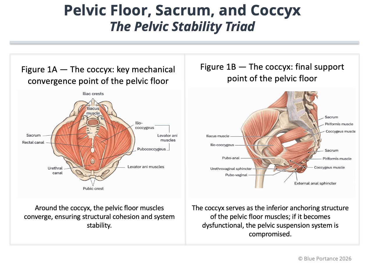
Muscular organization of the pelvic floor and role of the coccyx as a lower anchor point providing cohesion, stability, and support to the pelvic system (Standring, 2021; Vodušek, 2004).
3. Biomechanics: an indispensable intrinsic mobility
3.1 A little-known physiological mobility
Contrary to a widespread belief, the coccyx is not completely fixed. It has a limited but functional mobility, notably when sitting, during defecation, and during childbirth (Woon & Stringer, 2012; Nathan, Fisher & Roberts, 2010). This mobility allows the shape of the pelvis to adapt finely to mechanical stress and prevents pressure from abruptly concentrating on a single point (Maigne, Doursounian & Chatellier, 2000).
3.2 Mobility dysfunctions
The problem is therefore not mobility itself, but:
- first, excessive mobility (hypermobility, subluxation),
- or, conversely, restricted mobility, which prevents mechanical adaptation (Maigne, Doursounian & Chatellier, 2000).
A coccyx can look “normal” on static imaging while behaving pathologically as soon as the person sits down. This is why analyzing mobility (particularly under dynamic conditions) is key to understanding many persistent cases of coccydynia (Maigne, Doursounian & Chatellier, 2000; Nathan, Fisher & Roberts, 2010).
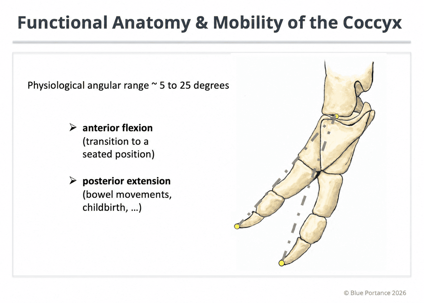
Physiological mobility of the coccyx (normal flexion ≈ 5–25°; beyond 25°, hypermobility) at the sacrococcygeal joint during changes in posture, contributing to the absorption and redistribution of stress in the seated position (Woon & Stringer, 2012; Maigne & Tamalet, 1996; Maigne, Doursounian & Chatellier, 2000).
4. A mechanical metaphor: the terminal articulated lever
Functionally, the coccyx can be compared to a small articulated lever located at the end of a large structure — the spine and the pelvis (Nathan, Fisher & Roberts, 2010).
- As long as this lever moves within a tolerance zone, it absorbs and distributes stress.
- If, on the other hand, it becomes too mobile, it moves out of its safety zone.
- Finally, if it becomes locked, stress can no longer be dissipated (Maigne, Doursounian & Chatellier, 2000).
Consequently, in both cases, every change of position — sitting down, standing up, leaning — places excessive stress on the surrounding tissues. The pain is then not mysterious: it is the logical consequence of a system that has lost its capacity to adapt (Maigne, Doursounian & Chatellier, 2000; Nathan, Fisher & Roberts, 2010).
5. The coccyx as a lower neurological crossroads
5.1 The ganglion impar and the sympathetic chains
The coccyx is not only a bony and muscular structure. On its anterior surface lies the ganglion impar (or ganglion of Walther), the terminal point of the two sympathetic chains. This center plays an important role in the transmission and modulation of pelvic pain (Sandrasegaram et al., 2020; Vodušek, 2004).
Chronic mechanical irritation of the coccyx (hypermobility, spicule, local conflict) can contribute to neurogenic sensitization, turning a localized mechanical discomfort into persistent pain, sometimes more widespread depending on the modulation and convergence pathways (Sandrasegaram et al., 2020).
5.2 Caudal continuity and modulation
In addition, the coccyx is indirectly connected to the spinal cord by the filum terminale, a caudal fibrous extension. This anatomical continuity suggests a potential role in transmission or modulation, although no direct causal link can be asserted on the basis of current data (Standring, 2021).
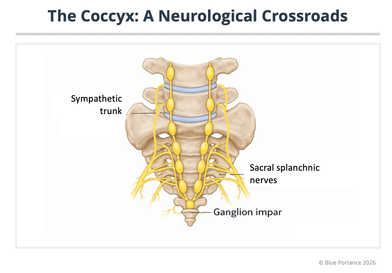
Position of the ganglion impar (Walther), termination of the sympathetic chains, and density of the pelvic network that may contribute to the modulation and persistence of tailbone pain (Sandrasegaram et al., 2020; Vodušek, 2004).
Interim conclusion — The “so what” of Page A
At the end of this page, a first reality becomes clear: the coccyx can be reduced neither to an anatomical vestige nor to a simple, isolated painful point. Despite its small size, it occupies a nodal position at the intersection of three inseparable dimensions: structural and muscular (support of the pelvic floor), biomechanical (indispensable intrinsic mobility), and neurological (lower crossroads of the pelvic autonomic nervous system) (Standring, 2021; Vodušek, 2004; Woon & Stringer, 2012).
This strategic position explains why damage to the coccyx — whether structural, functional, or resulting from their interaction — can have disproportionate clinical repercussions: stress concentration, disorganization of the pelvic sling, loss of adaptability, and neurological sensitization (Maigne, Doursounian & Chatellier, 2000; Sandrasegaram et al., 2020).
This page thus lays the explanatory foundation needed to address, in the following sections of the knowledge base: mechanical causes, the local vs referred pain distinction, structural vs functional coccydynia, the mechanisms of chronicity, and vulnerability factors (Nathan, Fisher & Roberts, 2010).
Scientific references
- Postacchini, F., & Massobrio, M. (1983). Idiopathic coccygodynia: Analysis of fifty-one operative cases and a radiographic study of the normal coccyx. Journal of Bone and Joint Surgery (American Volume), 65(8), 1116–1124.
- Maigne, J.-Y., Doursounian, L., & Chatellier, G. (2000). Causes and mechanisms of common coccydynia: role of body mass index and coccygeal trauma. Spine, 25(23), 3072–3079. doi: 10.1097/00007632-200012010-00015
- Maigne, J.-Y., & Tamalet, B. (1996). Standardized radiologic protocol for the study of common coccygodynia and characteristics of the lesions observed in the sitting position. Clinical elements differentiating luxation, hypermobility, and normal mobility. Spine, 21(22), 2588–2593. doi: 10.1097/00007632-199611150-00008
- Woon, J. T. K., & Stringer, M. D. (2012). Clinical anatomy of the coccyx: A systematic review. Clinical Anatomy, 25(2), 158–167.
- Nathan, S. T., Fisher, B. E., & Roberts, C. S. (2010). Coccydynia: a review of pathoanatomy, aetiology, treatment and outcome. Journal of Bone and Joint Surgery (British Volume), 92-B(12), 1622–1627. doi: 10.1302/0301-620X.92B12.25486
- Standring, S. (Ed.). (2021). Gray’s Anatomy: The Anatomical Basis of Clinical Practice (42nd ed.). Elsevier.
- Sandrasegaram, N., Gupta, R., & Baloch, M. (2020). Diagnosis and management of sacrococcygeal pain. BJA Education, 20(3), 74–79. doi: 10.1016/j.bjae.2019.11.004 (open access – PMC7807997)
- Vodušek, D. B. (2004). Anatomy and Neurocontrol of the Pelvic Floor. Digestion, 69(2), 87–92. doi: 10.1159/000077874
Doctrinal note
This knowledge base rests on the articulation between morphology and structural lesions (Postacchini & Massobrio, 1983), dynamic mobility and weight-bearing examination (Maigne, Doursounian & Chatellier, 2000), and precise anatomical attachments (Woon & Stringer, 2012; Standring, 2021). The neurogenic dimension is framed by the anatomical reality of the ganglion impar and the sympathetic pathways (Sandrasegaram et al., 2020), and by overall pelvic neuro-functional physiology (Vodušek, 2004), without extrapolating a direct causal link.
© Gil Ayache. The concepts, diagrams, terminology, and principles presented on this page constitute a work protected by copyright. They are made available to Blue Portance under an intellectual property license agreement, without transfer of economic rights or of authorship.
